


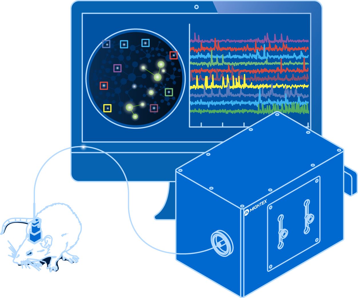
Calcium imaging enables neuroscientists to visualize the activity of large neuronal populations using fluorescent activity indicators (Grienberger & Konnerth, 2012). The invention of imaging technologies, such as the optical fiberscope or miniscope, allows for calcium imaging to be performed in freely-behaving animals with single-cell resolution (Ghosh et al. 2011). These capabilities enable neuroscientists to map the relationship between neural circuits and behaviour with single-cell resolution.
Despite many advantages, single-cell resolution calcium imaging systems are (1) more complex and generate large data sets that are difficult to handle, and (2) require more intricate surgical procedures. For labs just starting with calcium imaging or performing exploratory experiments, single-cell imaging may be too overwhelming or unnecessary.
Is there a simpler and less expensive technology for in vivo calcium imaging?
What is Fiber Photometry?
Fiber photometry, as a technology, enables the measurement of population-level activity with calcium imaging (Cui et al. 2014; Kim et al. 2016). Instead of visualizing individual neurons, the combined or ‘bulk’ activity signal from all neurons is measured from the neurons expressing the fluorescent activity indicators (Cui et al. 2014; Kim et al. 2016), which leads to various benefits elaborated below.
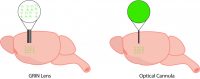
The lack of cellular resolution with fiber photometry is due to a simplified coupling mechanism between the brain and the imaging device. With an optical fiberscope or miniscope, a GRIN lens is implanted into the brain and this enables an image with cellular resolution to be collected from the brain (Resendez et al. 2016). Thus, the changes in an individual neuron’s activity can be detected. In comparison, fiber photometry uses a low-cost implanted cannula that does not provide cellular resolution for imaging but rather collects a cumulative and combined signal from all neurons.
Why Use Fiber Photometry?
Fiber photometry has become a very popular method for in vivo calcium imaging due to its many advantages.
First, fiber photometry has a low entry barrier for labs wanting to integrate calcium imaging into their experiments. Compared to single-cell resolution imaging technologies, fiber photometry provides a much simpler data output, lower data output, and has a lower associated cost.
Second, from a technical and biological standpoint, fiber photometry is easier to implement, compared to other calcium imaging technologies. Due to the use of a cannula, it requires a less invasive surgery, similar to that of optogenetics. Fiber photometry also provides very sensitive and fast imaging by using a photodetector, compared to a camera, even though the latter may be employed for multi-region experiments (this will be discussed in more detail in the following post).
Third, fiber photometry has advantages for specific applications:
- With no miniscope on the head of the animal and smaller data files, fiber photometry is more suitable for long freely-behaving experiments.
- Smaller data files make it a more suitable imaging technique for exploratory experiments.
- Fiber photometry can be used to image multiple brain regions or multiple subjects simultaneously (Kim et al. 2016).
- Fiber photometry has been used to image fluorescent indicators beyond calcium, such as neurotransmitter fluorescent activity indicators (Leopold et al. 2019).
- Optogenetics can be easily integrated for all-optical experiments to probe neural circuits (Kim et al. 2016).
There are two main disadvantages of fiber photometry for calcium imaging, which may or may not be crucial depending on your experiment. First, the resolution of fiber photometry is limited to population-level imaging and cannot be used to visualize individual neurons. Second, incorporating optogenetics in a fiber photometry system is limited to population-level stimulation and cannot be used to deliver cellular-resolution optogenetics to manipulate individual neurons.
Next Post
What Equipment Do You Need to Perform Fiber Photometry?
——————————
Related Products for Fiber Photometry
Fiber Photometry
and optogenetics to probe complex neuronal networks.
- Simultaneous Calcium Imaging and Optogenetics
- Multi-Region Investigation
- Reconfigurable Platform for Single-Cell Resolution Imaging
- High-Quality Imaging with Scientific Cameras