For many bioscience applications such as optogenetics, precisely-timed and high-intensity light pulses are required to activate channelrhodopsins (ChR2, ChR1 etc.) and halorhodopsins (NpHR), in order to excite and/or inhibit neurons using a microscopy light source. Other applications, such as Calcium imaging or voltage imaging, call for light sources which offer not only powerful and stable illumination, but also fast response time and long lifetime. Mightex has developed a wide range of market-leading BioLED sources for such applications.
Key features:
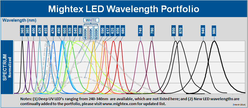
- Wide range of wavelengths from UV, visible, NIR to white
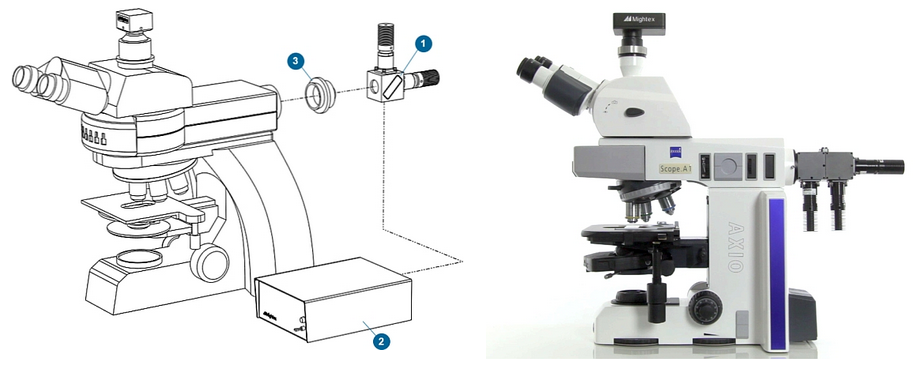
- Collimated beam, readily compatible with microscope’s infinity optical path
- High-power solid state light sources
- Long lifetime and high stability
- Fast response time, with modulation frequency up to 100KHz
- Beam combiners for combining up to 8 LEDs into one beam
- Adapters for most microscope makes (Olympus/Nikon/Zeiss/Leica etc)
- Optional LED controllers with manual, analog, or TTL/software controls
- Compact, machined metal housing with integrated heat sink
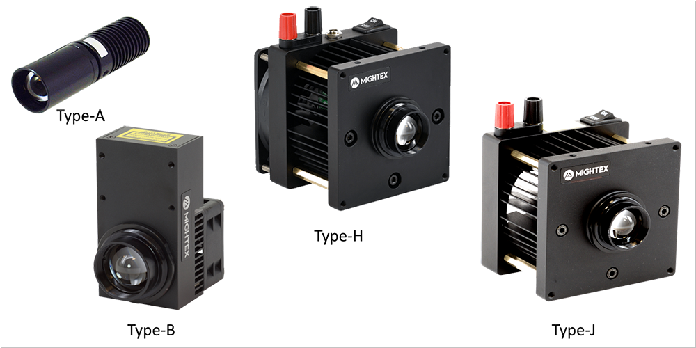
LCS-Series Microscope LEDs, Single Wavelength
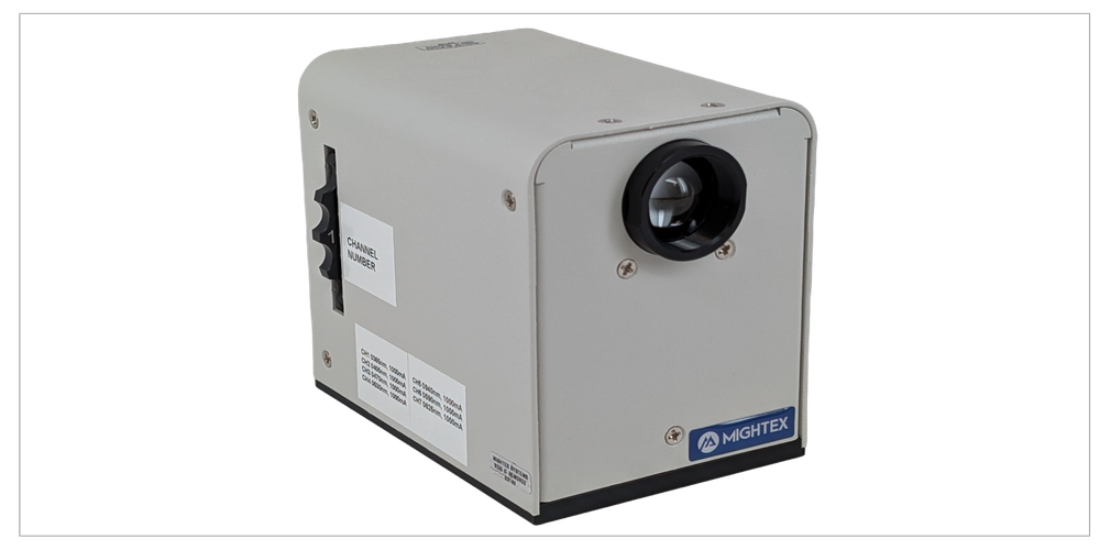
WheeLED Wavelength-Switchable LED for Microscopes
Mightex microscope LED light sources are modularized fully-customizable turn-key solutions for optogenetics, fluorescence excitation, and other biophotonics applications. Precisely-timed and high-intensity light pulses are required in optogenetics experiments to activate channelrhodopsins (ChR2, ChR1 etc.) and halorhodopsins (NpHR) in order to excite and inhibit neurons using a microscopy light source. To meet these requirements, Mightex has developed a proprietary “IntelliPulsing” technology to allow BLS-series sources to output significantly higher power in pulse mode than what the LEDs are rated for in CW mode.
Features:
- High-power UV/VIS/NIR/white microscope LED light sources
- Interchangeable fiber with SMA connector
- No moving parts in optical path
- Multiple mounting features for lab and OEM applications
- Optional LED controllers
- Compact, machined metal housing with integrated heat sink
- Locking electrical connector
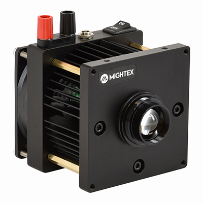
2. Select LED Configuration
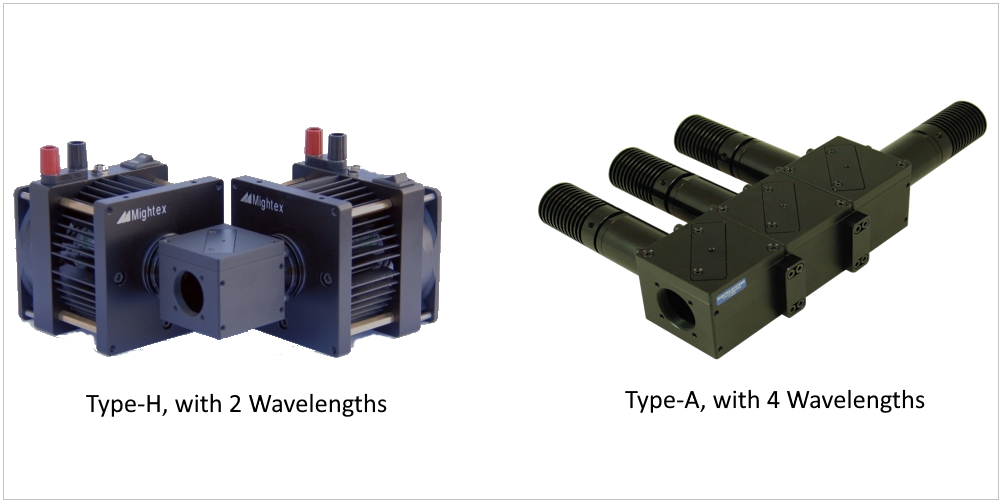
Single-Wavelength Microscopy Light Sources
Type A, Type B, Type J & Type H
Multi-Wavelength Microscopy Light Sources
Type A, Type B, Type J & Type H
Wavelength-Switchable Microscopy Light Source
Type A
3. Select LED Controller
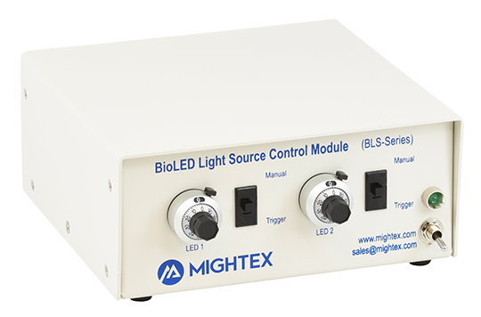
Manual/Analog Controllers for Microscope LED Light Source
BLS-1000-2, BLS-3000-2, BLS-13000-1, and BLS-18000-1
Manual/Software/Analog/TTL Controllers for Microscope LED Light Source
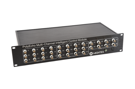
Add a PolyEcho Multi-Channel Intelligent Control Module (PEC-CM12-U)
4. Microscope Coupling
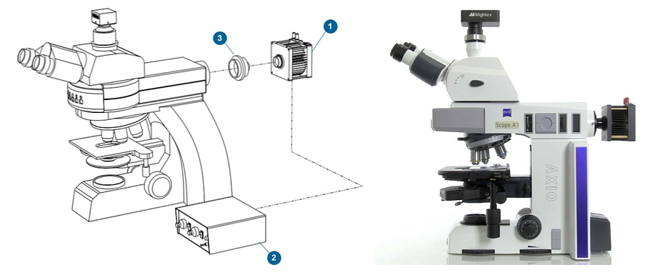
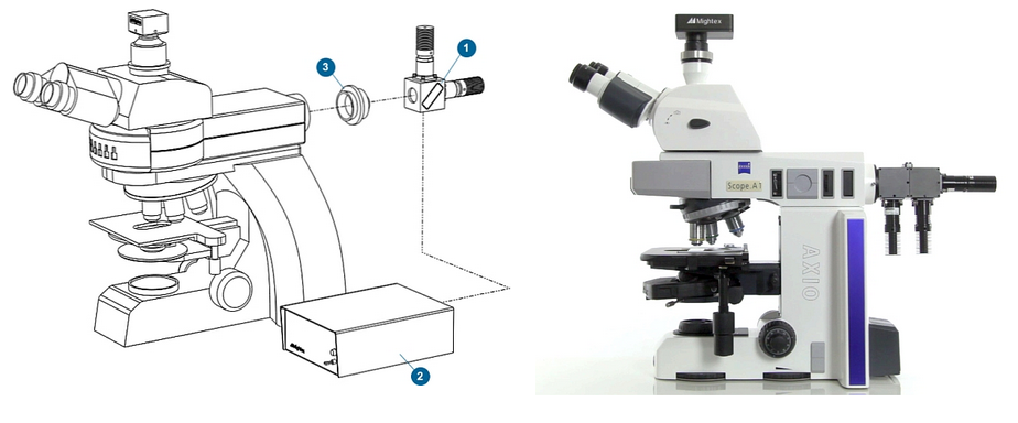
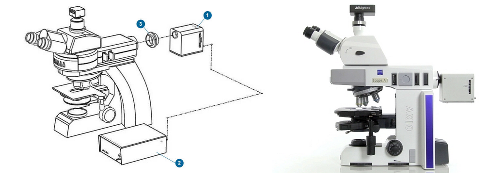
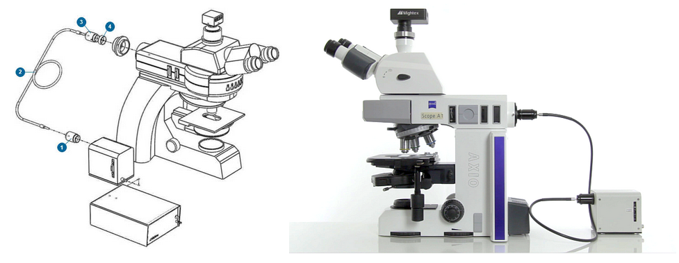
Microscope adapters are available for all major manufacturers (Nikon, Olympus, Leica, Zeiss) to couple microscope LED sources. If you have a different microscope, please contact us.
Direct-Coupling
Lightguide-Wavelength
Microscopy LED Application Examples
Abdelfattah et al., developed novel chemigenetic indicators for in vivo voltage imaging in neurons. To illuminate their indicators, the group used Mightex’s Microscope LEDs.
Crandall SR, Patrick SL, Cruikshank SJ, & Connors BW
In this work, Crandall et al., used Mightex’s Microscope LEDs to optically stimulating different neuronal pathways connecting to pyramidal neurons found in L6 of the whisker somatosensory cortex of mice.
To study phase resetting curves in vivo, Higgs and Wilson used Mightex’s Microscope LEDs to establish whether their novel method of optogenetic barrage stimulation would work during extracellular spike recordings (a recording method often used in in vivo experiments).
Microscopy LED Inquiry Form
Fill out the form below and we will contact you!
"*" indicates required fields


