

?")
CRACM, or channelrhodopsin-2(ChR2)-assisted circuit mapping, is an optical method used to dissect large, complex neural circuits with optogenetics (Petreanu et al. 2007).
To start, ChR2 is expressed in the neuron population of interest, enabling optogenetic activation of this specific cell-type. While recording from an individual neuron using electrophysiology, patterned light stimulation is employed to scan the field of view, activating neurons expressing ChR2. Any activated neurons that are connected to the recorded neuron will elicit an electrophysiological response, and these responses can then be used to generate a heatmap of connections between the recorded neuron and surrounding neurons.
Why Use CRACM?
Two other methods used to test the connections between neurons are paired-recordings and uncaging. Paired recordings are used to test the connections between multiple neurons with high precision; however, they are limited in the number of neurons that can be recorded from and the range (e.g. across multiple layers)(Kissinger et al. 2020). On the other hand, uncaging can be used for examining connections across a long-range; however, it lacks cell-type specificity (Kissinger et al. 2020). Therefore, the current circuit mapping techniques have certain limitations, which can be addressed using CRACM.
CRACM has three main advantages compared to the existing techniques mentioned above. First, by using optogenetics, the expression of ChR2 can be restricted to specific cell-types. This enables the examination of connections with specific cell-types rather than unknown cell-types. Second, CRACM can be used at a lower magnification, enabling scientists to examine a large field of view across multiple layers and a larger cell-population. While the existing techniques are limited to local neural circuits, CRACM can be used to investigate long-range projections. Lastly, with technological advancements, CRACM can be limited to specific layers, the size of spot scanned across the layers, and which neurons are illuminated/stimulated. As a result, CRACM is a powerful tool enabling scientists to investigate large and complex neural circuits.
What Do You Need to Perform CRACM?
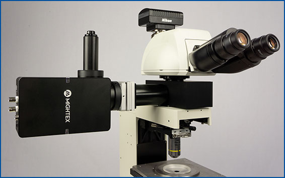
First, you will need a microscope and the required electrophysiology equipment. This will be used for (1) the electrophysiological recordings and (2) synchronization between the optogenetic stimulation and recording using TTL triggering.
Second, you will need a digital micromirror device (DMD), such as Mightex’s Polygon DMD pattern illuminator. Compared to a standard light source that illuminates the entire field of view and, hence, stimulates all neurons indiscriminately, a DMD pattern illuminator enables scientists to target any specific regions of interest (ROI’s) on the sample, such as a designated neuron or a specified group of neurons. Thus, a DMD pattern illuminator is a crucial component for CRACM, as it enables targeted light to be scanned across the sample for optogenetic stimulation. As an example, Mightex’s market-leading Polygon DMD pattern illuminator can be easily integrated into any microscope for optogenetic stimulation and synchronized with external electrophysiology equipment.
Lastly, a light source (such as an LED or a laser) is required to couple light through the DMD pattern illuminator, in order to optogenetically stimulate the sample. The light source provides the light/power, whereas the micromirrors in the DMD pattern illuminator decides, through software, precisely where light is exposed on the sample. Mightex’s Polygon pattern illuminator is highly flexible and it can be used with a multitude of light sources, such as LEDs and lasers. For most optogenetic experiments, an LED will be adequate; however, when a wider field of view is required, a laser source may be necessary for optogenetic stimulation in order to provide sufficient optical power.
Examples Publications using Mightex’s Polygon DMD Illuminator for CRACM
Kissinger et al. (2020). Visual Experience-Dependent Oscillations and Underlying Circuit Connectivity Changes are Impaired in Fmr1 KO Mice. Cell Reports.
Kissinger et al. 2020 published a paper in Cell Reports examining perceptual deficits in a mouse model of Fragile X syndrome (Fmr1 knockout mice). They investigated any potential connectivity changes in V1 by performing CRACM using Mightex’s Polygon DMD Illuminator. Using this technique, the authors were able to examine single-cell connections between cells using optogenetics.
Channelrhodopsin was expressed in control and Fmr1 knockout animals by crossing them with Thy1-ChR2-YFP mice. Naive or experienced animals were sacrificed, and patch-clamp electrophysiology was used to test connections between cells. Specifically, connections between layer 5 – layer 4 and layer 5 – layer 5 cells were tested. While cells were patched, individual cells were stimulated in a 10 by 10 grid (0.67 mm x 0.67 mm) using the Polygon (covering a square from L2/3 to L5) at a 10X . A 470 nm LED light source with 10 ms optogenetic light pulses through a 10x objective was used (0.3 mW under the objective) to stimulate individual neurons.
Kissinger et al. showed a significant potentiation in layer 5 to layer 4 fast-spiking cells in control animals, but no change in Fmr1 knockout animals following perceptual experience. In addition, a significant depression in layer 5 to layer 5 neurons was seen in Fmr1 knockout animals following perceptual experience. This study demonstrates significant differences in how an animal model of Fragile X syndrome encodes information. As well, using Mightex’s Polygon for CRACM to demonstrate associated circuit changes in Fmr1 knockout mice.
Other Publications
- Galloni et al. (2021). Dendritic domain-specific sampling of long-range axons shapes feedforward and feedback connectivity of L5 neurons. BioRxiv.
- Anastasiades et al. (2020). Mediodorsal and ventromedial thalamus engage distinct L1 circuits in the prefrontal cortex. Neuron.
- Andrasi et al. (2017). Differential excitatory control of 2 parallel basket cell networks in amygdala microcircuits. PLoS Biology.
- Shimizu & Stopfer (2017). A population of projection neurons that inhibits the lateral horn but excites the antennal lobe through chemical synapses in Drosophila. Frontiers in Neural Circuits
——————————
Related Products for CRACM
for Cellular-Resolution Optogenetics
spatiotemporal control of light with subcellular resolution.
- Cellular-Resolution Optogenetics
- Simultaneous Multi-Region Illumination
- Synchronize with Electrophysiology or Imaging
- Compatible with Any Microscope