


Current advanced imaging techniques, such as calcium imaging, can record cortical activity with high spatial precision and cell-type specificity using the expression of fluorescent sensors and implantation of a cortical window; however, this technique can be quite invasive and complex.
For head-fixed experiments measuring general changes in cortical activity in different areas, there is not always a need for such complex imaging techniques, but rather a simplistic imaging method is more suitable to examine basic physiology.
Is there a more simplistic imaging method available?
What is Intrinsic Optical Imaging?
Intrinsic optical imaging (or intrinsic signal imaging) is an imaging method that enables scientists to indirectly record neural activity non-invasively by measuring hemodynamic changes in the brain (Morone et al., 2017).
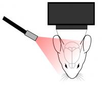
In a typical setup, a mesoscope is used to image the cortex of an animal, and an external light source is used to shine light onto the cortex off which light will then be reflected. The camera on the mesoscope detects slight changes in reflected signal off the cortex, which is indicative of neural activity (Hillman et al., 2008)(what components are needed for intrinsic imaging will be discussed in the next post). These changes are quite small (1% change), and they are measured relative to a baseline control (Morone et al., 2017).
Specifically, intrinsic optical imaging exploits the differential spectral absorption properties of oxygenated and deoxygenated hemoglobin (Hillman et al., 2008). Where the typical response around the site of neural activity shows an increase in deoxygenated hemoglobin and blood flow followed by an increase in oxygenated hemoglobin (Juavientt et al., 2018). These changes can be detected by using different wavelengths for total hemoglobin and blood flow (500-599nm) and deoxyhemoglobin (600-699nm) measurement (Morone et al., 2017). Thus, most intrinsic imaging experiments use multiple wavelengths to detect different components in the hemodynamic response related to neural activity.
In cortex-wide or cortical imaging experiments, intrinsic optical imaging is commonly used to measure changes in neural activity due to a behaviour or presentation of a stimulus. The relative temporal and spatial precision of intrinsic imaging is normally appropriate for these types of experiments (Juavientt et al., 2018).
Why Use Intrinsic Optical Imaging?
A common reason neuroscientists employ intrinsic optical imaging—compared to other more advanced imaging techniques (e.g. calcium imaging)—is to image neural activity in large cortical areas and examine basic physiology (Juavientt et al., 2018). For these applications, intrinsic optical imaging provides both high enough spatial (100um) and temporal resolution (1-2 seconds to peak) to map stimulus-evoked cortical activity in a head-fixed animal (Lu et al., 2017).
One advantage of intrinsic optical imaging is being able to image neural activity with minimal invasiveness and no expression of fluorescent reports. Due to the simplicity of this imaging technique, intrinsic optical imaging does not necessitate the implantation of a cortical window to replace the skull or thinning of the skull (Juavientt et al., 2018); however, if combined with other imaging techniques, such as two-photon imaging, a cortical window would be necessary. Using the reflected properties of hemodynamic changes in the brain provides a convenient way to image neural activity without the need for fluorescent reporters to be expressed (Hillman et al., 2008). These two factors, in combination with minimal equipment, makes intrinsic imaging a fairly inexpensive tool to probe neural activity across large cortical regions.
With such ease of use, there are potential pitfalls associated with intrinsic imaging, and these are mostly dependent on the application being studied. First, due to the inherent properties of the brain, there is light scattering associated with intrinsic imaging and thus, this method is limited to superficial regions of the cortex (Hillman et al., 2008). Second, intrinsic optical imaging cannot provide single-cell or cell-type specific imaging. This limitation is due to the spatial resolution of the technique and solely relying on hemodynamic changes, which cannot provide cell-type specific changes (Juavientt et al., 2018). Thus, intrinsic optical imaging is more useful as a general activity reporter across the cortex. Third, depending on the cortical events being studied or behavioural stimuli, intrinsic imaging may be too slow to measure changes in cortical activity (Lu et al., 2017). Intrinsic optical imaging reflects changes over a matter of seconds, and this may not be ideal for certain experiments.
To overcome the limitations of intrinsic optical imaging, it would be appropriate to implement or complement these experiments using more advanced techniques such as calcium imaging, voltage imaging, or voltage sensitive dyes. In particular, calcium imaging is a well-suited and established imaging technique for resolving single-cells or cell-type specific activity and measuring neural activity with higher temporal precision.
Next Post
What Components Do You Need for Intrinsic Optical Imaging?
——————————
Related Products for Intrinsic Optical Imaging
Cortex-Wide Intrinsic Optical Imaging
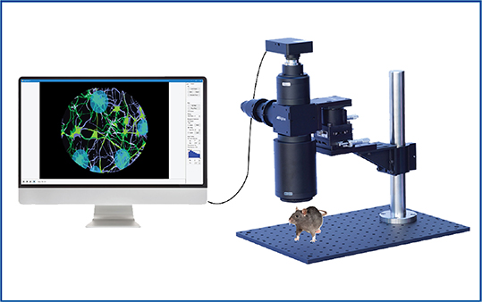
targeted optogenetics, and calcium imaging.
- Large Field of View for In Vivo Imaging and Optogenetics
- Targeted Optogenetics
- Reconfigurable Mesoscope
- Designed for In Vivo Experiments