Overview
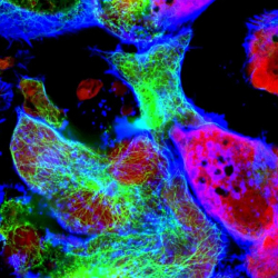
Spatial biology investigates the structural organization of cells and molecules within intact tissues, enabling researchers to study how cellular function is influenced by location and context.
By employing patterned illumination such as Mightex’s market leading Polygon DMD Patterned Illuminator, scientists can dynamically and precisely control light exposure to image, or manipulate biomolecules across defined tissue regions. High-resolution, light driven techniques, integrated with spatial omics platforms, enhance the ability to visualize and manipulate molecular signals in complex tissues.
For more details, please click here ➡️ eBook: Patterned Illumination for Spatial Biology
Key System Requirements
Central to spatial biology technologies are imaging-based approaches and other imaging-integrated methods that profile genes, transcripts, proteins, and metabolites across spatial dimensions and biological scales. These techniques often operate across diverse tissue types and spatial resolutions, requiring high flexibility and precision. While specific workflows using patterned illumination may have unique demands, most share the following core requirements:

- High precision and customizability, allowing free-form regions of interest (ROIs) with complex shapes
- Supports multiplexed ROI selection
- Compatibility with multiple wavelengths
- Submicron resolution
- Non-destructive cell type targeting
- Easy integration with a variety of microscopes
Polygon DMD Patterned Illuminator
Mightex’s market leading Polygon DMD Patterned Illuminator is designed and developed to meet these challenging requirements. With a DMD array of more than 1 million individual pixels, it achieves a large projection field-of-view, without compromising resolution– enabling precise and non-destructive light delivery to even the most intricate tissue regions. It supports multiplexed ROI selection and is fully compatible with a large wavelength spectrum (350 nm – 1000 nm). Seamlessly integrable with most microscope systems and supported by intuitive pattern design and control software, the Polygon offers a powerful and flexible solution for advancing spatial biology research.

Customer Success and Publications

High-depth spatial transcriptome analysis by
photo-isolation chemistry
This study demonstrates a photo-activated approach using Mightex’s Polygon DMD Patterned Illuminator, enabling deep transcriptome profiling from precisely defined regions–ranging from tissue domains to subcellular structures. The method reveals spatially localized gene expression with with high sensitivity and resolution, advancing capabilities of spatial biology to explore even the smallest functional units within multicellular organisms.
Dr. Mizuki Honda & Prof. Yasuyuki Ohkawa
Kyoto University, Japan & Kyushu University, Japan
Using spatial transcriptomics integrated with Mightex’s Polygon DMD patterned Illuminator, this study compared neuroblasts migrating toward cortical injury sites within the ventricular-subventricular zone (V-SVZ). The analysis revealed distinct gene expression patterns, including reduced proliferation and enhanced migration-related signaling, influenced by local TGF-𝛽 activity near the lesion.
Takuya Miyamoto & Prof. Kazunobu Sawamoto
Nagoya City University, Japan


Light-Seq: Light directed in situ barcoding
By combining in situ imaging with spatially targeted barcode attachment, researchers were able to sequence specific cell populations based on location, morphology, or protein markers. The researchers used Mightex’s DMD Patterned Illuminator to illuminate a group of ROIs with UV light in the presence of complementary docking sequences to label wit a particular fluorescence tag.
Jocelyn Y. Kishi & Prof. Peng Yin
Harvard Medical School, USA