

")
Selective Optogenetic Stimulation with MEA to Detect Spread – Papasavvas et al. 2020 eNeuro
Interneurons are directly connected by gap junctions to other interneurons, and this provides a highly specific excitatory link. It is relatively unknown how gap-junction mediated propagation may be linked to increased activity. Papasavvas et al. 2020 tested the hypothesis that raised extracellular potassium levels may be associated with extreme levels of neuronal activity.
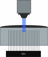
Papasavvas et al. 2020 used Mightex’s Polygon DMD Illuminator to illuminate only specific electrodes on the MEA.
To test this hypothesis, the authors recorded electrophysiological activity in slices with normal or increased potassium levels, and they used optogenetics to activitate parvalbumin-expressing interneurons with ChR2. Field potentials were measured using a MEA placed along layer V of the occipital cortical slices. Mightex’s Polygon DMD Illuminator was used to illuminate three to four adjacent electrodes (300-400um x 10 um square) with a 470nm LED. The experiments were performed under a 4x objective, yielding an approximate 2 mw/mm2 of intensity under the objective. Following optogenetic stimulation, the spread of activity was measured and sampled at 100um spaces between individual electrodes in conjunction with pharmacological manipulations.
Papasavvas et al. 2020 showed at baseline extracellular potassium levels that neural activity was limited to the electrodes illuminated. However, by increasing potassium levels, there was increased spontaneous activity and a time-locked burst of firing at least 150um away from recording sites following optogenetic stimulation. The proportion of slices that showed propagation from the illumination site increased with the associated extracellular potassium levels. In this paper, Papasavvas demonstrate using patterned illumination and MEA recordings that extracellular potassium levels affect the spontaneous activity of interneurons.
Soma-Targeted Optogenetics with Electrophysiology – Anastasiades et al. 2020 Biorxiv
There are reciprocal connections between the prefrontal cortex (PFC), mediodorsal (MD), and ventromedial (VM) thalamic nuclei. However, how these different thalamic nuclei communicate with the prefrontal cortex is unclear. Anastasiades et al. 2020 set out to understand the nature of these connections using techniques such as anatomical tracing, electrophysiology, and optogenetics.
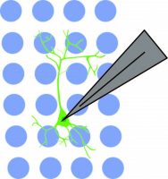
Anastasiades et al. 2020 used Mightex’s Polygon DMD Illuminator to optogenetically stimulate individual interneuron somas while recording from a pyramidal neuron.
The authors first demonstrated using anatomical tracing that the VM projections were found in layer 1a of the PFC and MD projections in layer 1b of the PFC. Specifically, it was found that NDNF+ cells receive inputs from VM in layer 1a of the PFC, and VIP+cells receive inputs from MD in layer 1b in MD. These findings suggested separate PFC-thalamic circuits.
With these direct connections established, Anastasiades et al. 2020 investigated how these different cell types—that receive direct input from the thalamus—communicate within local PFC circuits. They recorded responses from local parvalbumin interneurons, somatostatin interneurons, and pyramidal neurons following optogenetic stimulation of NDNF+ and VIP+ cells that expressed ChR2. Interestingly, VIP-evoked IPSCS occurred only in somatostatin interneurons, but not pyramidal cells or parvalbumin interneurons. Conversely, NDNF+-evoked IPSCS occurred in parvalbumin interneurons and pyramidal cells, but not somatostatin interneurons. These findings extended upon the author’s previous experiments by demonstrating distinct microcircuits within the PFC.
The authors showed that the connection between NDNF+ cells and pyramidal cells was not limited to PFC Layer 1. Soma-targeted optogenetics was used to identify the location of NDNF+ cells connected to pyramidal cells. NDNF+ cells expressed optogenetic construct st-Chrome, which was activated by blue light. Mightex’s Polygon DMD illuminator with a 473nm LED was used to illuminate a 10 x 10 pseudo-random grid (mapping area of 750um x 750um) under a 10x objective. Subsequent responses were measured in pyramidal cells using electrophysiology.
First, the authors established the parameters for st-Chrome soma-targeted optogenetic activation. Activation was only detected when the soma was directly illuminated, and no response was detected when the illumination spot was 75um from the soma. The authors showed using soma-targeted optogenetics that pyramidal cells receive input from NDNF+ predominantly in Layer 1, but also receive some input from Layers 2/3 and 5. In this paper, Anastasiades et al. 2020 show an intricate long-range PFC-thalamic circuit.
References
- Papasavvas CA, Parrish RR, & Trevelyan AJ. (2020). Propagating Activity in Neocortex, Mediated by Gap Junctions and Modulated by Extracellular Potassium. eNeuro, 7(2).
- Anastasiades PG, Collins DP, & Carter AG. (2020). Mediodorsal and Ventromedial Thalamus Engage Distinct L1 Circuits in the Prefrontal Cortex. BioRxiv.
——————————
Related Products for Optogenetics
for Cellular-Resolution Optogenetics
spatiotemporal control of light with subcellular resolution.
- Cellular-Resolution Optogenetics
- Simultaneous Multi-Region Illumination
- Synchronize with Electrophysiology or Imaging
- Compatible with Any Microscope