PHOTOPATTERNING WITH MERCAPTOPROPYLSILATRANE AND THE POLYGON 1000
In our study, we use the Mightex Polygon1000 to photopattern molecules, such as fluorescent dyes, on silanized surfaces. Silanes form self-assembled monolayers on glass, and by choosing silanes with different tailgroups, we obtain different surface functionalities. Here, we use thiol tailgroups, and project UV (365 nm) patterns on the glass substrate, while it is submerged in a solution of photoinitiator and alkyne-terminated molecules. The light catalyzes the thiol-yne “click” reaction to attach the alkyne-functionalized molecules to the surface.
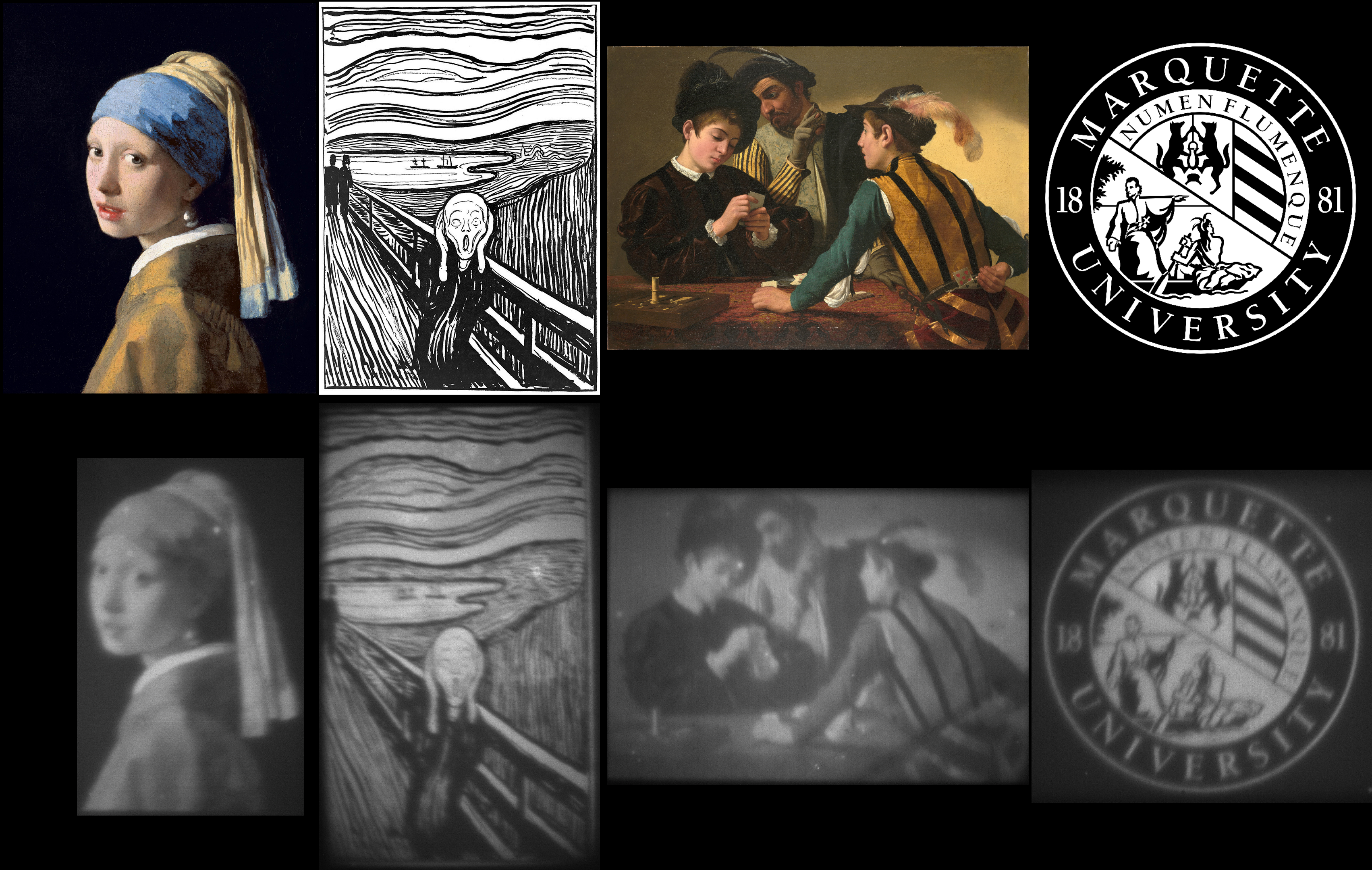
By projecting different patterns, we control where molecules are attached, and so obtain monolayers of arbitrary shape. We can even obtain monolayers with partial coverage – by breaking down a grayscale image into a series of different black and white images, we can individually control the exposure time at each pixel, and correspondingly tune the density of deposited dye molecules at each position. We have demonstrated this process with two different dyes, 3-ethynyl perylene and FAM alkyne. We have also used substrates coated with different silanization agents: the commonly used mercaptopropyltrimethoxysilane, and mercaptopropylsilatrane, a molecule previously reported to form smoother monolayers.
We are currently pursuing the ability to photopattern appropriately functionalized gold nanoparticles in an arbitrary gradient using this technique. Our long-term interest is to create surface-based chemical reaction networks, which interact with the environment and conduct multi-component reactions on surfaces. For this purpose, we need to selectively localize different molecules in specific locations, creating the need for developing the photopatterning capability detailed here.

Figure 1. The top row contains the original patterns, and the bottom row displays the captured fluorescence images from the fluorescein derivative dye (FAM alkyne, 5-isomer) that was attached to the patterned location, observed with 455 nm light. From left to right, the images are: 1) “Girl with a Pearl Earring” by Johannes Vermeer, 2) black-and-white version of “The Scream” by Edvard Munch, 3) “The Cardsharps” by Michelangelo Merisi da Caravaggio, and 4) the seal of Marquette University. Each image in the figure has an area of only 0.48 mm^2.

Author: Mark Mitmoen
Bio: Mark is a PhD student in the Nanomaterials Lab in the Department of Chemistry at Marquette University. He is supervised by Dr. Ofer Kedem.


