
Serotonergic modulation of vigilance states in zebrafish and mice


Published on 2024/04/19 Research powered by Mightex’s Polygon1000 ![]()
![]()

Introduction
In the animal kingdom, behavior is intricately linked to internal brain states, which are influenced by neural dynamics affecting sensation, cognition, and action (1, 2). Animals adapt their behaviors dynamically to suit their circumstances, requiring the brain to generate appropriate internal states beforehand. One crucial internal state involves selectively enhancing the sensitivity of certain sensory inputs while neglecting others, particularly important for survival, especially in response to potential threats like predators (3, 4). Vigilance behavior, characterized by reduced locomotion and heightened alertness, enables animals to swiftly detect and avoid danger, although it may impact forging efficiency and group dynamics.
The 5-HT neurons in the dorsal raphe nucleus (DRN) have a significant role in the serotonergic tone to the forebrain and consecutively shaping behavior states. Despite the functional diversity of these neurons and the complex activation of postsynaptic receptors, the precise mechanism by which the DRN 5-HT system modulates neural dynamics to control internal brain states remains uncertain. While the activation of the ascending norepinephrine system in wakeful mice prompts transitions between internal states, studies indicate that DRN 5-HT neurons function oppositely, inducing locomotor reduction or even stationary states in response to various behavioral challenges in zebrafish and mice. The article presents evidence suggesting that an unconventional 5-HT-driven innate mechanism at the circuit level shapes the internal brain state underlying vigilance behavior, shedding light on a mechanism operating across species.
Methods
In this study the researchers employ a combination of behavioral assays, neuroimaging techniques, genetic manipulation, pharmacological interventions, and optogenetic stimulation to investigate the neural mechanisms underlying vigilance behavior in zebrafish and mice.
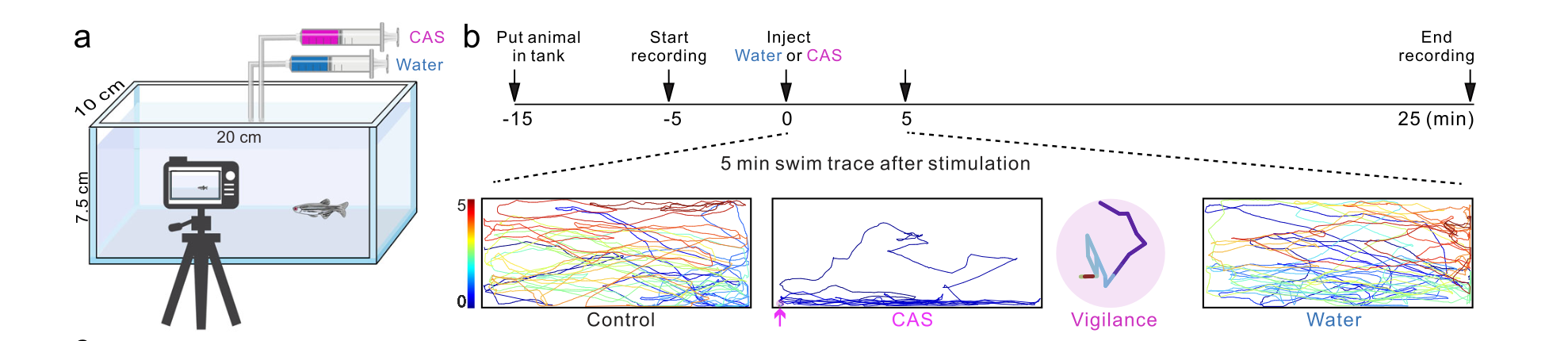
Figure 1: Illustration of experiment setup and procedures showing zebrafish swimming patterns before and upon water or CAS addition
The DRN 5-HT neurons in Zebrafish are selectively targeted using Mightex’s Polygon1000 for optogenetic stimulation. Overall, these techniques provide insights into the neural circuitry and molecular mechanisms underlying vigilance behavior across species.
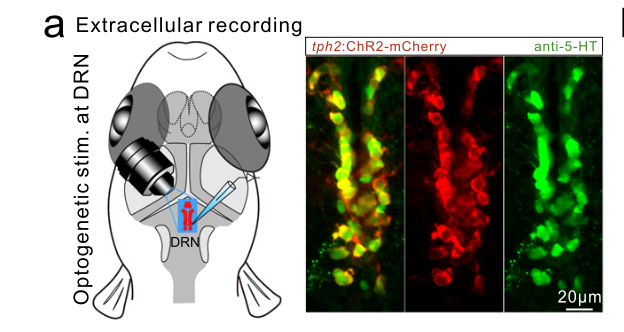
Figure 2: Illustration of optogenetic stimulation combined with extracellular recording of DRN 5-HT neurons in zebrafish
Results
The study investigated the optogenetic modulation of vigilance behavior in zebrafish and mice. In zebrafish, they observed that conspecific alarm substance (CAS) administration induced a persistent vigilance behavior characterized by reduced locomotion and increased sensitivity to aversive stimuli. This behavioral state persisted even when fish were placed in a new tank with fresh water, indicating its robustness. Neural activity imaging revealed synchronized neuronal oscillations in the dorsal pallium, particularly in the central zone (Dc region), after CAS treatment. This synchronized state correlated with the persistence of the vigilance behavior and was dependent on the activation of DRN 5-HT neurons.
Further experiments demonstrated that the intense activation of DRN 5-HT neurons induced by CAS treatment led to increased release of serotonin in the Dc region, which was crucial for maintaining the synchronized neuronal state and the vigilance behavior. Specifically, the synchronized neurons in the Dc region were identified as glutamatergic neurons expressing the 5-HT7a receptor. Activation of these receptors triggered burst firing patterns in glutamatergic neurons, leading to synchronized oscillations. Optogenetic stimulation of DRN 5-HT neurons reliably induced and maintained the vigilance behavior, further confirming the role of serotonin in regulating this internal brain state.
The study also extended its findings to mice, showing a similar mechanism of vigilance behavior modulation mediated by DRN 5-HT neurons projecting to the prefrontal cortex. Optogenetic activation of these neurons in mice induced synchronized neuronal activity and reduced locomotion, mimicking the vigilance behavior observed in zebrafish.
The persistence and intensity of DRN 5-HT neuron activation were critical for generating and maintaining this synchronized state, highlighting an evolutionarily conserved mechanism across species.
These findings provide valuable insights into the neural basis of vigilance behavior and its modulation through optogenetic manipulation of serotonin pathways.
References:
Anderson, D. J. Circuit modules linking internal states and social behaviour in flies and mice. Nat. Rev. Neurosci. 17, 692–704 (2016).
Lee, S. H. & Dan, Y. Neuromodulation of brain states. Neuron 76, 209–222 (2012).
Oken, B. S., Salinsky, M. C. & Elsas, S.M. Vigilance, alertness, or sustained attention: physiological basis and measurement. Clin. Neurophysiol. 117, 1885–1901 (2006).
Hart, T. et al. Sparse and stereotyped encoding implicates a core glomerulus for ant alarm behavior. Cell 186, 3079–3094 (2023).
Megha Patwa, Applications Specialist at Mightex
To read the full publication, please click here.