


Activation of Somatostatin-Expressing Neurons in the Lateral Septum
Improves Stress-Induced Depressive-like Behaviors in Mice
Published on 2022/12/14 Research powered by Mightex’s OASIS Fiberscope 


Check out this recent publication using the OASIS Fiberscope! The paper outlines research by Huanhuan Li and colleagues from Geoff Lau’s lab at the City University of Hong Kong.
Introduction
The limbic system, including the lateral septum (LS), plays a crucial role in mood disorders, such as depression. However, the role of the LS in depression remains unclear. A potential important role is ascribed to neurons expressing somatostatin (SST) in particular. Li and colleagues hypothesized that LS SST neuronal activity is linked to stress and depression.
Methods & Findings
In order to examine the contributions of SST-expressing neurons of the LS in depression-like behaviors in mice, Li and colleagues leveraged calcium imaging using the Mightex OASIS Fiberscope and GCaMP6s sensor in SST-cre mice. Their findings show that LS SST neuronal activity was significantly reduced in two mouse models of depression (Chronic Restraint Stress [CRS] and repeated lipopolysaccharide [LPS] injections).
In order to determine whether artificial manipulation of LS SST neuronal activity could attenuate the depression-like behavioural observations, Li and colleagues combined their calcium imaging with optogenetics to excite LS SST neurons during depression model experiments. The authors found that optogenetic excitation of LS SST neurons produced antidepressant-like effects in the LPS depression model. Taken together these findings elucidate the LS SST neurons as a potential therapeutic target for the treatment of depression.
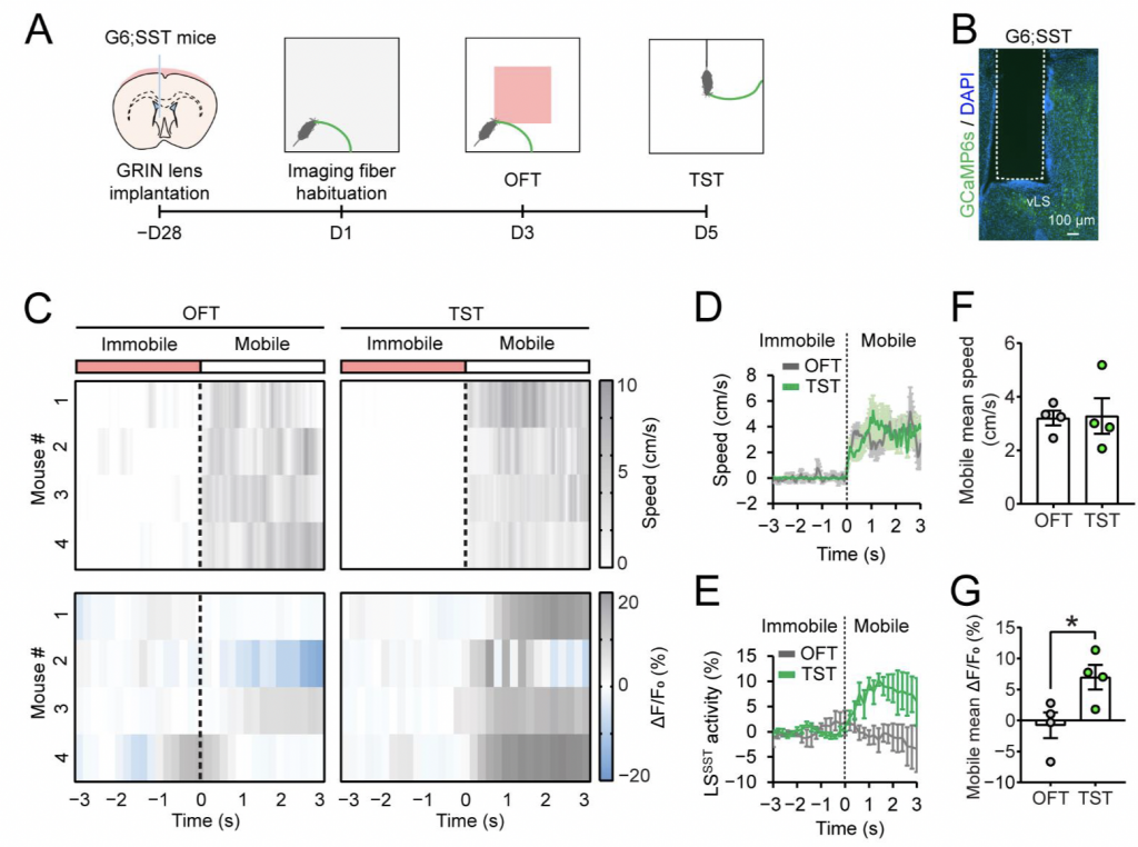
Figure 1: Adapted from Source
Panel A. The surgical and experimental preparation stages.
Panel B. GRIN lens location within LS.
Panel C. Calcium imaging results. Change in fluorescence depicted for 4 mice; changing in fluorescence (bottom) and corresponding behavioural changes (top) seen as gradient overtime for Open Field Test (OFT; left) and Tail Suspension Test (TST; Right)
Panels D & F. Behavioural quantification for OFT and TST; the dotted line represents the initiation of the mobile phase.
Panels E & G. Calcium imaging quantification for LS SST neurons during OFT and TST; the dotted line represents the initiation of the mobile phase.
Findings and Conclusion
The above described experiments demonstrate the capabilities of the Mightex OASIS Fiberscope for calcium imaging in freely moving mice. To learn more about using the system for your research, get in touch today!
Catherine Thomas, PhD Senior Liaison and Development Scientist at Mightex
To read the full publication, please click here.