OPTOGENETIC CHARACTERIZATION OF MUSCULAR RESPONSES IN THE TIBIALIS ANTERIOR (TA) MUSCLE

Peripheral optogenetics is a technique that allows motor nerves to be stimulated with light after using a viral vector to induce expression of light sensitive ion channels, or opsins, along the nerve. This new alternative to electrical stimulation for reanimation of paralyzed muscles provides muscle specific stimulation, a more natural muscle fiber recruitment order, and reduced muscle fatigue compared to traditional electrical stimulation [1], [2].
Along with these advantages of optical stimulation, there are many challenges to successfully implementing this technique, such as a potential immune response to the injected virus and a long viral expression pathway from injection site to the spinal cord. All these factors affect the temporal and spatial pattern of opsin expression in the desired nerve, making knowledge of the expression pattern crucial for effective optical stimulation.
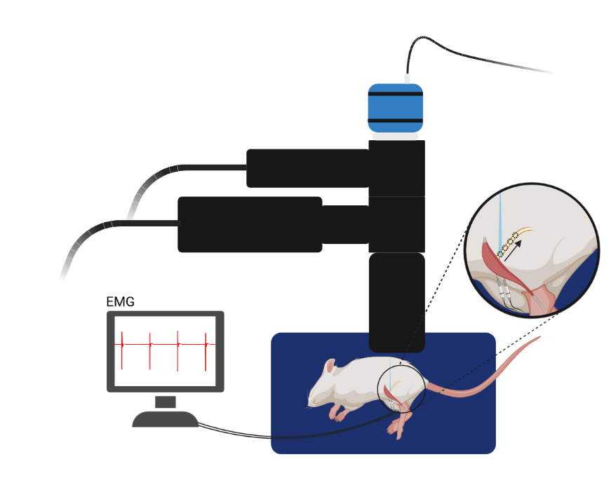
Figure 1. Experimental Schematic
Video 1. Pattern optogenetic stimulation of TA Muscle in vivo.
In this video, we used the Polygon system to stimulate the peroneal nerve in small segments starting from the distal muscle insertion moving proximally towards the sciatic nerve in a rat expressing Channelrhodopsin-2 (ChR2) in the peroneal nerve after an intramuscular virus injection in the Tibialis Anterior (TA) muscle. At the same time, we recorded muscle response to the optical stimulation in the TA muscle with intramuscular electromyography (EMG). This experiment allows us to characterize the strength of opsin expression and muscle responses along the length of the nerve at any given time point, which we have found to be highly variable after virus injection. The Polygon system also allows us to assess the spatial pattern of opsin expression along the nerve, both along its length as well as its width. This will serve to give us an even better idea of how opsin expression varies spatially in the nerve and enable us to activate specific muscle and nerve fibers.
[1] C. Towne, K. L. Montgomery, S. M. Iyer, K. Deisseroth, and S. L. Delp, “Optogenetic Control of Targeted Peripheral Axons in Freely Moving Animals,” PLoS ONE, vol. 8, no. 8, p. e72691, Aug. 2013, doi: 10.1371/journal.pone.0072691. [2] M. E. Llewellyn, K. R. Thompson, K. Deisseroth, and S. L. Delp, “Orderly recruitment of motor units under optical control in vivo,” Nat. Med., vol. 16, no. 10, pp. 1161–1165, Oct. 2010, doi: 10.1038/nm.2228.
Author: Emma Moravec
Bio: Emma is a Biomedical Engineering PhD student in the Neural Engineering, Interfacing, Modulation, and Optimization (NEIMO) Lab in the Joint Department of Biomedical Engineering at Marquette University and the Medical College of Wisconsin. She is supervised by Dr. Jordan Williams.


