

")
We are pleased to share the recent work of Zubayer Ibne Ferdous, a doctoral student in the Berdichevsky Lab at LeHigh University and a recent recipient of the Mightex Travel Award for their work using the Mightex Polygon for patterned optogenetics examination of neural computation in cortical culture neurons. Their findings demonstrate a new method of integrating spatiotemporal dynamics into the study of neural network dynamics.
Introduction
Confined neural networks in vitro with optical interface can be used to classify spatiotemporal information. Studies showed that cortical microcircuits can be intrinsically responsive to spatiotemporal patterns no matter how they are connected. We found that the dissociated cortical network with an optical interface could process spatiotemporal information from the distributed input patterns and store their information in the output neurons in real time.
Ibne Ferdous et al. obtained dissociated cortical cultures from postnatal day 0-1 rat pups. Before culturing cells, they established confinement (area ~1 𝑚𝑚^2 and ~4 𝑚𝑚^2) using polydimethylsiloxane (PDMS) structures. After 1 day of plating cells, they were co-transfected with ChannelRhodopsin 2 (ChR2) and jRGeco1a for simultaneous optical patterned stimulation and optical recording. The authors used Polygon 400-G (Mightex) to achieve targeted optical stimulation to input patterns.
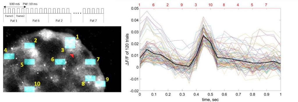
Figure 1: Stimulation patterns and parameters using the Mightex Polygon.
Optical response (Right panel) of one output neuron with input patterns at different locations. (Top left) Schematic of input patterns. (Bottom left) locations of inputs and one output (red arrowhead).
Experimental Design
Network bursts represent chaotic activation and do not carry information about the input. First, the authors designed an input stimulation protocol to suppress bursts. The results showed significant reduction of bursts in confined networks when distributed inputs were optically stimulated and each input pattern was repeated at least once a second. Increases in Ca2+ baseline level of all neurons was correlated with successful burst suppression. In the absence of population bursts, there was a strong correlation between outputs and input patterns that indicates processing of spatial information (Fig.1). To perform the experiment, Ibne Ferdous and colleagues divided the total working area of the Polygon into 10X10 grids (Fig. 1), and 20X20 grids (Fig. 2), then assigned one grid to one pattern (Fig. 1) out of 100 grids and four grids to one pattern (Fig. 2) out of 400 grids. Each pattern was then activated 5 times at 50 Hz with 10 msec pulse width. In Fig. 1, a sequence of 10 patterns was generated at a random order and recorded optical activity from one random output neuron (outside the stimulated grids; shown in red arrowhead). The authors observed strong response in the output neuron with pattern 3 and a weaker response with pattern 1.
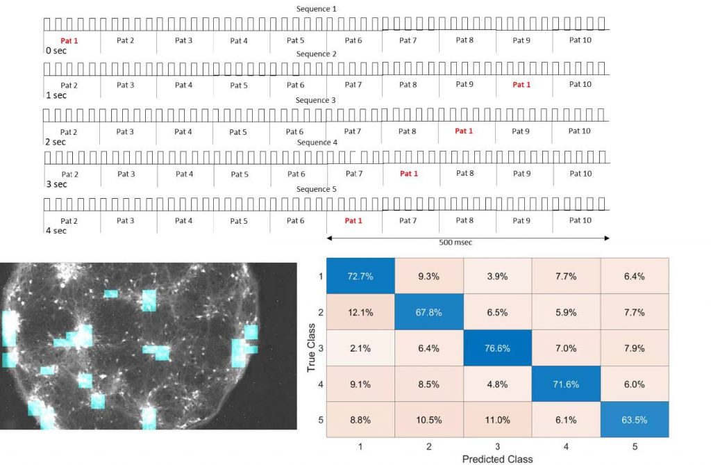
Figure 2.Classification accuracy of 5 sequences based on the sequence end responses from all outputs. Here, only position of pattern 1 in each sequence was moved. Ibne Ferdous and colleagues then recorded the responses of pattern 10 for all outputs. These recorded datasets (80% training – 20% testing) were used to classify 5 sequences (bottom right).
Further classification of spatiotemporal input patterns allowed authors to check how long the network could store information of those patterns. They designed the stimulation protocol for this experiment by generating five sequences of 10 patterns where the final pattern in these five sequences was fixed and rest of the patterns were circularly shifted to left by 100 msec in each sequence (Fig.2 top panel). Optical activities were then recorded from all output neurons while running these five sequences for 120 trials.
These recorded datasets from all the outputs (80% training – 20% testing) were used to classify 5 sequences using linear discriminant analysis. The results showed that the cortical network in vitro could classify and store information of spatiotemporal patterns up to 500 msec (Fig. 2 bottom right).
Conclusion
These findings demonstrate that patterned optical stimulation in a confined network in vitro can be a useful tool to study network dynamics. By applying distributed optical stimulation patterns in the network, the authors suppressed population bursts and at the same time used those patterns to classify spatiotemporal information in the output neurons. These experiments outline a paradigm of reservoir computing with living neural network. This model can further be used to study toxicity or influences of commercial drugs on spatiotemporal information in neural networks.