
How to combine opsins and fluorescent sensors to minimize crosstalk
using the OASIS Fiberscope
using the OASIS Fiberscope


A central goal of behavioural neuroscience is to understand connections between brain activity and behavioural outcomes in order to elucidate neural circuits underpinning particular behaviours and to highlight putative targets for therapeutic approaches in disease models. With the goal in mind, observing neural activity, or proxies thereof, in freely behaving animals provides one vital tool. In order to then examine the precise role that a particular neural circuit or node plays in a behaviour, neuroscientists must then manipulate the circuit in question and observe any marked change in behaviour as a result. One popular way to do this is by combining in vivo freely behaving one photon calcium imaging – to observe calcium fluctuations in real time as a proxy for neural activity- with targeted optogenetics to augment activity of a neuronal population of interest. Together, these two techniques combined with behavioural observation allow neuroscientists a unique approach to connecting brain activity to behaviour in real time. However, both techniques rely on transmission of particular wavelengths of visible light to excite opsin or sensors with high temporal and spatial precision. Often, the wavelengths needed for each technique overlap resulting in a problem known as crosstalk.
Minimising cross talk between wavelengths is critical when combining cellular resolution one photon calcium imaging and targeted optogenetics, as both modalities often rely on overlapping spectral bands. A primary strategy is careful spectral planning: selecting calcium sensors and opsins with maximally separated excitation and emission spectra, and pairing them with narrow-band excitation sources and high-quality dichroic mirrors and emission filters. In Mightex’s OASIS Fiberscope system, which supports multi-wavelength illumination through a shared fiber bundle, using well-matched bandpass filters at both the illumination and detection paths helps suppress bleed-through from optogenetic stimulation light into the calcium imaging channel (see Figure 1). Additionally, minimizing excitation power to the lowest level that still provides adequate signal-to-noise reduces unintended activation or fluorescence from off-target wavelengths.
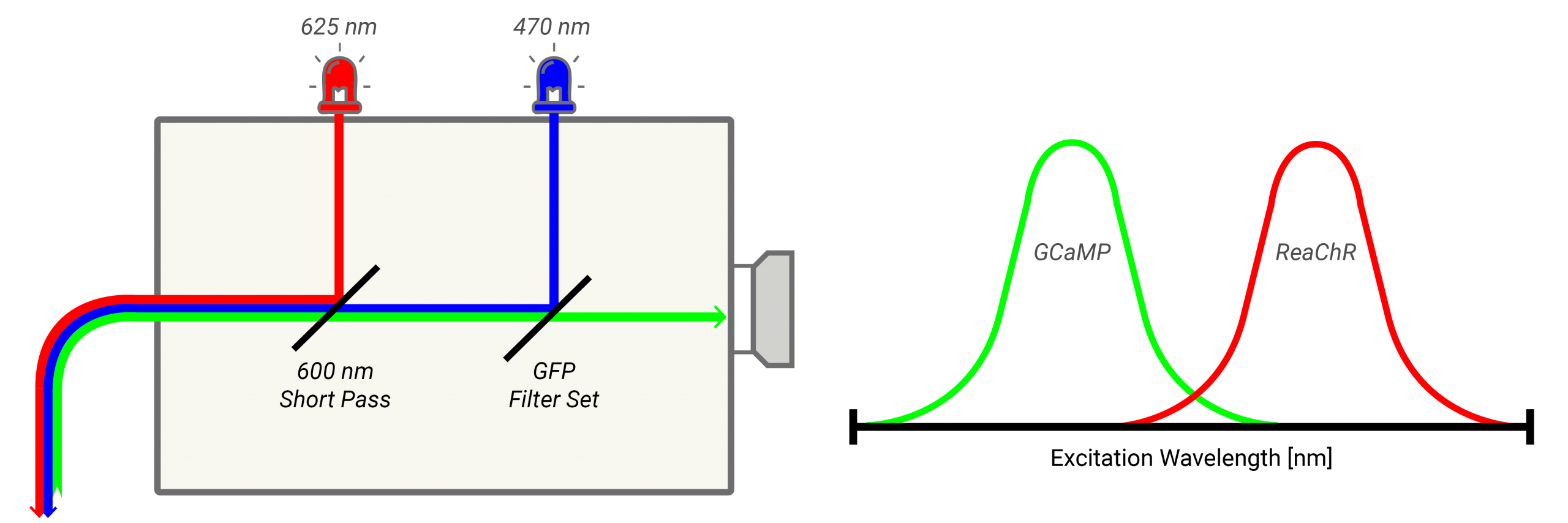
Figure 1.
Temporal separation further reduces cross talk in one-photon fiber photometry, cellular resolution imaging and optogenetic stimulation (see Figure 2a). Alternating calcium imaging and optogenetic stimulation in time—either through fast wavelength switching or interleaved stimulation–acquisition cycles—allows fluorescence signals to be recorded when optogenetic light is off, eliminating direct optical contamination (see Figure 2b). The OASIS fiberscope’s precise control over LED timing and intensity can be leveraged to synchronize illumination with camera exposure. Finally, post-hoc computational approaches, such as reference channel subtraction using control recordings, can correct residual cross talk that remains after optical and temporal optimization, resulting in cleaner calcium signals and more reliable interpretation of neural activity during optogenetic manipulation.

Figure 2.
To learn more about the OASIS Fiberscope and its features, click here or contact our customer support team.