


of refractive error in the mouse retina
Published on 2024/01/30 Research powered by Mightex’s Polygon1000 ![]()

So Chung Him, & Dr Pan Feng, Exploration of the mechanism of refractive error in the mouse retina. The Hong Kong Polytechnic University, (2023).


We use the Mightex Polygon to investigate the biophysical properties of retinal ganglion cells (RGCs) under myopic and astigmatic conditions. To simulate myopia, the retina was stretched using tractional forces. Astigmatic blur images were projected onto the retina to study the effects of astigmatism. The hypothesis is that the elongation of the eyeball in myopia and the creation of an optical aberration pattern in astigmatism causes changes in the image projected onto the retina, which can impact the signals sensed by retinal ganglion cells.
To explore the biophysical responses of retinal ganglion cells response to focused/defocused images under stretching, as well as the effects of astigmatic component light (different directions of light bar) image projection on the mouse retina. Our laboratory utilizes the Mightex Polygon1000 system to provide various image stimuli in conjunction with the patch clamp whole-cell recording.
The Polygon allows us to stimulate RGCs’ receptive field with focused sinusoidal grating by projecting to the photoreceptor layer and defocused grating by projecting out of the photoreceptor layer (Figure 1A). We utilized the four directions of the light bar and spots/annulus pattern as stimuli to test the polarity and light response of neurons (figure 1B).
To test whether the biophysical properties of RGCs being stretched, including soma stretching and retina stretching to mimic myopia elongation (Figure 1C) can still reflect focused/defocused stimuli and whether RGCs’ somata being twisted, partly mimic astigmatic light to RGC (Figure 1D), alter their response properties.
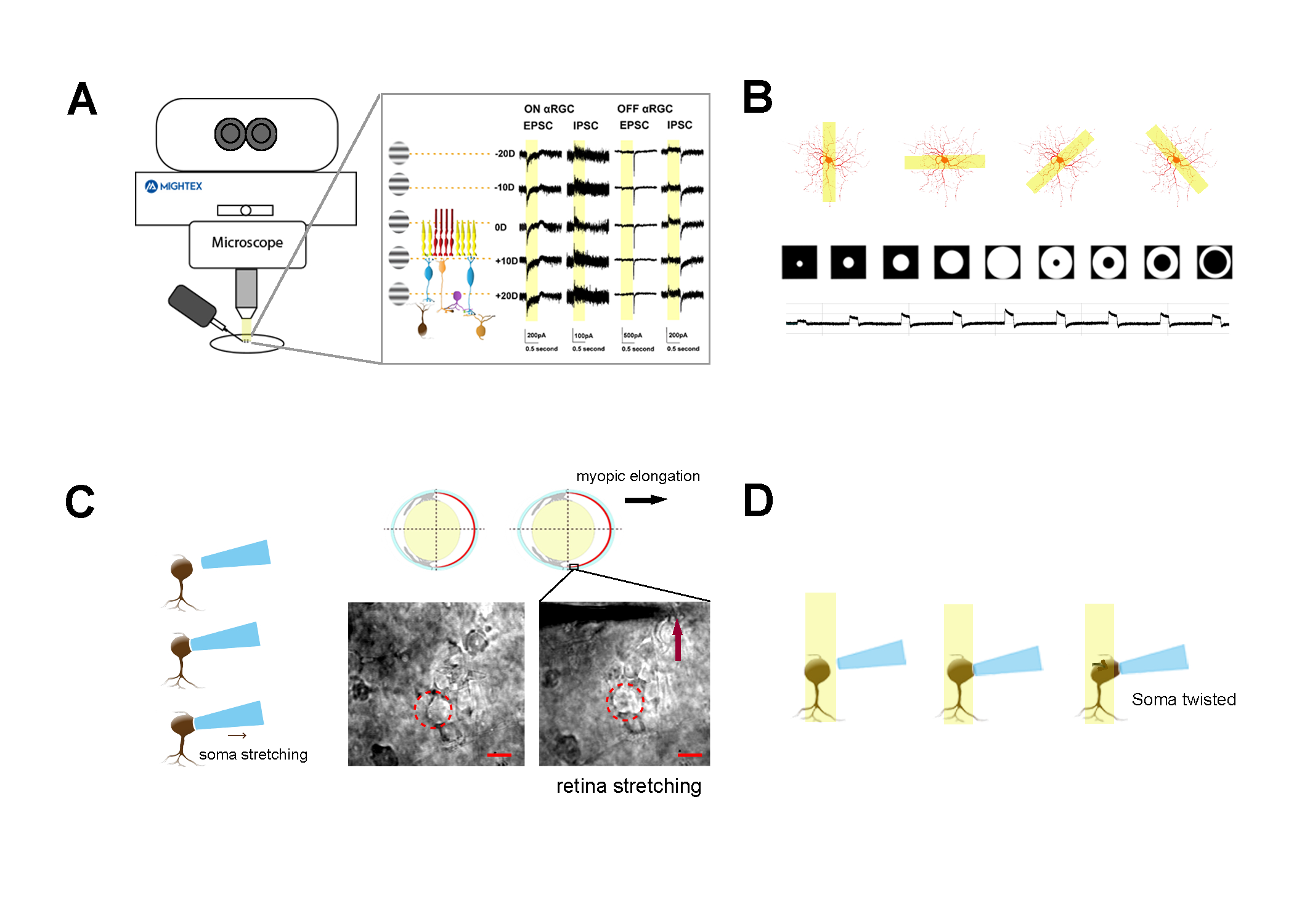
Figure 1. Using a digital micro-mirror device (Polygon1000, Mightex) and patch clamp systems to study the biophysical properties of retinal ganglion cells under both normal and myopia/astigmatism conditions. A) A sinusoidal grating project to the photoreceptor layer defines as 0D (focus). 100 μm defocus would be expected to generate plus or minus 20 diopters of refractive error under microscopy depending on the direction of defocus. B) A 20µm x 200µm light bar in four directions and various spots/annulus generated by the Mightex Polygon1000 system projected onto neuron’s receptive fields and the responses of spots/annulus. C) The soma of αRGCs and retina were stretched by tractional forces to mimic myopia status, followed by the same stimuli’s projection as A suggest. D) αRGC somas were twisted using a suction pipette to orient a projected 30-degree angle light.
With the aid of Mightex Polygon1000, we were able to generate different types of grating patterns and better explore the biophysical properties of specific neurons. We have analyzed αRGCs response to specific sinusoidal grating under normal and stretching somas/retinas. Our conclusion is that αRGCs can reflex the projection of focus/defocus stimuli even under myopic conditions, supporting the hypothesis that myopia development is driven by visually-guided and retinas(RGCs) play a crucial role. Additionally, the hypothesis of whether “retinal astigmatism” exists is controversial. The astigmatic experiment design is the first step in our project, we decomposed the astigmatic blur into different directions of light bar and twisted cell soma (soma relative to light angle change from astigmatic blur). We also irradiated different angles of laser light and inserted Plano-cylindrical lenses between grating projections to induce party or fully astigmatic blur. The result is that αRGCs can reflex these different stimuli.
References:
- Schaeffel F. Test systems for measuring ocular parameters and visual function in mice. Front Biosci. 2008;13:4904-4911. Published 2008 May 1. doi:10.2741/3049
- Wienbar S, Schwartz GW. The dynamic receptive fields of retinal ganglion cells. Prog Retin Eye Res. 2018;67:102-117. doi:10.1016/j.preteyeres.2018.06.003
- Kee CS. Astigmatism and its role in emmetropization. Exp Eye Res. 2013;114:89-95. doi:10.1016/j.exer.2013.04.020
Author: So Chung Him
Bio: PhD student in the School of Optometry at The Hong Kong Polytechnic University.