


and application to developing ferret cortex
Published on 2024/01/29 Research powered by Mightex’s Polygon1000 ![]()

Mulholland, H. N., Jayakumar, H., Farinella, D. M., & Smith, G. B., All-optical interrogation of millimeter-scale networks and application to developing ferret cortex. Journal of Neuroscience Methods, 403, 110051 (2024).


The intricate neural networks underlying sensory perception and behaviour span across various brain regions. Optical imaging advancements have transformed the study of these networks, offering broad data collection with high spatial resolution. Simultaneously, optogenetics allows precise manipulations within these networks.
To address the challenge of integrating large-scale imaging and manipulation, the researchers developed an “opto-macroscope.” This novel system enables targeted optogenetic manipulations with high spatial and temporal precision across the brain while concurrently capturing calcium imaging data.
Using the opto-macroscope, the study successfully reactivated endogenous cortical networks in the developing ferret visual cortex, showcasing its potential for investigating and manipulating functional modules identified through optical imaging.
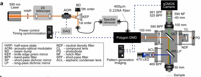
Figure 1: Schematic of the opto-macroscope light path. A 590 nm laser (orange line) passes through a power-control AOM and enters the Polygon DMD device for patterned stimulation. Simultaneously, GCaMP excitation light (blue line) and its emission (green line) are directed towards the sample using specific dichroic mirrors. To prevent unwanted stimulation light from reaching the camera, various filters are employed.
The opto-macroscope was designed to enable precise optogenetic manipulations and high-sensitivity neural imaging across a 2D brain surface. GFP-based calcium sensors, like GCaMP6 and jGCaMP8, were paired with red-shifted optogenetic tools such as Chrimson to minimize cross-talk between imaging and optogenetic stimulation. Imaging employed a back-illuminated sCMOS camera and a tandem-lens macroscope for optimized light collection over a 4 mm imaging field of view. Optogenetic stimulation was facilitated by the Mightex Polygon1000-DL DMD device, allowing precise spatial targeting of neural activity patterns. Control of stimulation parameters and patterns was managed via custom Python-based software interfaced with the DMD and other optical components.
The opto-macroscope presented offers a novel method to investigate neural networks on a larger scale, spanning millimeters, by combining optical imaging with targeted optogenetic manipulation using the Polygon DMD device. While based on prior designs, this system introduces several improvements, including narrow spectral bandwidth for reduced photodamage and flexibility for various red-shifted optogenetic probes. In combination with the Polygon’s precise and controlled optogenetic targeting, the opto-macroscope’s versatility suggests potential applications across various species and brain regions, facilitating deeper insights into neural network dynamics and behaviours.
Megha Patwa, Applications Specialist at Mightex
To read the full publication, please click here.