

")
Targeted Illumination in Zebrafish – Tabor et al. 2018 Current Biology
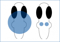
Tabor et al. 2018 examined the cellular-basis of sensory filtering in zebrafish using prepulse inhibition (PPI). PPI is performed by pre-exposing an animal to a mild stimulus, and following this they are presented with an alarming stimulus. A natural response to PPI is a reduction in response to an alarming stimulus due to the pre-exposure.
Tabor et al. 2018 selectively activated hindbrain neurons or R4 neurons in zebrafish.
In their initial experiments, calcium imaging was used to determine that the R4 domain in the zebrafish brain correlated with a PPI response. The authors decided to test whether they could simulate this PPI response by selectively activating the R4 domain in Zebrafish with optogenetics.
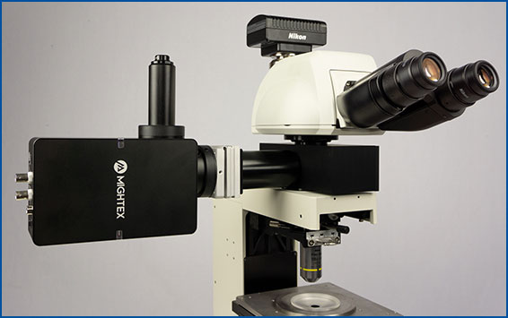
Using Mightex’s Polygon DMD Illuminator, Tabor et al. 2018 optogenetically stimulated individual regions in the zebrafish brain expressing ChR2 and measured the PPI response. The Polygon with a 460 nm LED was used to illuminate a 341 um wide square across the hindbrain or smaller regions using 33-50 um wide squares to selectively activate R4 neurons.
Optogenetic stimulation of either the hindbrain or R4 neurons suppressed the startle response when the pulse was presented 400ms after illumination. This response was not inhibited when neurons fired during the pulse-alone or before the stimulus. In this paper, Tabor et al. 2018 demonstrate a brainstem motor circuit for PPI in zebrafish to test sensory gating.
Targeted Illumination with Patch-Clamp Electrophysiology – Andrasi et al. 2017 PLOS Biology
Andrasi et al. 2017 investigated the connectivity between pyramidal neurons and interneurons in the basal nucleus of the amygdala and how these interactions regulate activity. They explored these questions using a variety of methods including optogenetics, patch-clamp electrophysiology, and microscopy techniques.
Andrasi et al. 2017 selectively activated surrounding pyramidal cells to test connections with interneurons.
In one experiment, the authors tested whether cholecystokinin (CCK) and parvalbumin (PV) interneurons receive direct input from pyramidal neurons in the amygdala. Andrasi et al. tested this by optogenetically stimulating surrounding pyramidal neurons that expressed ChR2 while recording the activity of CCK or PV interneurons with electrophysiology. By using Mightex’s Polygon DMD Illuminator, Andrasi et al. were able to illuminate individual cells using 15-20 um spots for 50ms of blue light on randomly chosen pyramidal cells surrounding the interneurons. The post-synaptic response following optogenetic stimulation was measured in CCK or PV cells.
The authors demonstrated using cellular-resolution optogenetic stimulation that pyramidal cells were more likely to be connected to PV interneurons than CCK cells. In addition, using the XY coordinates of the record cell and distance from the optogenetically stimulated pyramidal soma, they created a connectivity map. They demonstrated that PV cells were connected to closer pyramidal cells, rather than long distance, and CCK connections were not dependent on distance. In conclusion, this paper showed different functions for PV and CCK interneuron cell networks in amygdala.
References
- Tabor KM et al. (2018). Presynaptic Inhibition Selectively Gates Auditory Transmission to the Brainstem Startle Circuit. Current Biology, 28(16), 2527-2535.
- Andrasi et al. (2017). Differential Excitatory Control of 2 Parallel Basket Cell Networks in Amygdala Microcircuits. PLOS Biology, 15(15).
Next Post
How Can Patterned Illumination be Used in Optogenetics Experiments? (Part 3)
——————————
Related Products for Optogenetics
for Cellular-Resolution Optogenetics
spatiotemporal control of light with subcellular resolution.
- Cellular-Resolution Optogenetics
- Simultaneous Multi-Region Illumination
- Synchronize with Electrophysiology or Imaging
- Compatible with Any Microscope