
Patterned Optogenetics Meets Cellular-Resolution Calcium Imaging:
Advancing In Vivo Behavioural Neuroscience
with Mightex Polygon and OASIS Fiberscope
Advancing In Vivo Behavioural Neuroscience
with Mightex Polygon and OASIS Fiberscope



Discover how combining the Mightex Polygon Patterned Illumination System with the OASIS fiberscope enables precise optogenetics, improved calcium imaging, and closed-loop neuroscience experiments.
Modern behavioural neuroscience seeks not only to observe neuronal activity but also to connect the activity of specific neuronal populations to particular behaviours. While genetically encoded calcium indicators, such as GCaMP, have made it possible to monitor the activity of hundreds of neurons simultaneously in freely behaving animals, conventional optogenetic stimulation often remains comparatively coarse (Given and Sparta, 2017). Whole-field illumination activates every opsin-expressing neuron within the illuminated region, making it difficult to isolate the contribution of individual neurons or functional ensembles (Carter and De Lecea, 2011). Furthermore, in experiments where multi-brain region imaging and optogenetics is combined, it is challenging with traditional optogenetic approaches to selectively target individual regions for circuit-specific manipulation
By combining the Mightex Polygon Pattern Illuminator for targeted optogenetics with the OASIS Fiberscope for cellular resolution imaging, researchers can integrate high-resolution multi-region calcium imaging with spatially selective optogenetic stimulation in a single experimental workflow. Instead of illuminating an entire field of view, Polygon projects dynamic illumination patterns that can selectively target individual neurons or user-defined groups of neurons identified through OASIS imaging. This combination enables more precise interrogation of neural circuits while preserving the flexibility required for freely behaving experiments.
Neural circuits are rarely homogeneous. Even neighboring neurons can encode different sensory inputs, behavioural states, or motor outputs. Furthermore, seemingly homogenous neuronal populations receive varied afferents that can differently sculpt the activity of neuronal subpopulations to encode any number of features (Groenewegen et al., 1999). Consequently, stimulating an entire population may mask the contribution of the specific neurons responsible for a given behaviour.
Patterned illumination overcomes this limitation by allowing researchers to define regions of interest (ROIs) directly from calcium imaging data. Polygon’s DMD-based illumination can then project light only onto selected neurons or functional ensembles while leaving surrounding neurons unstimulated.
This capability enables experiments such as:
Such an approach opens the door to previously impossible experimental designs, and improves experimental specificity, providing greater confidence in conclusions when linking neuronal activity to behavioural outcomes. Rather than asking whether a brain region is involved in a behaviour, researchers can begin asking which cells within that region are responsible.
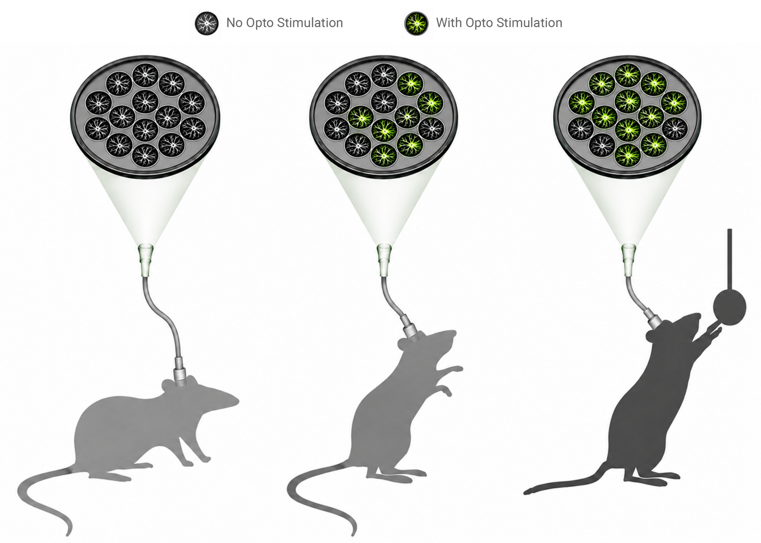
Figure 1. Example OASIS Fiberscope + Polygon experimental workflow.
One of the practical challenges of combining one photon calcium imaging with optogenetics is maintaining high-quality fluorescence measurements during optical stimulation. Broad illumination can introduce background excitation, increase scattered light, and create optical crosstalk that reduces imaging quality.
Spatially targeted optogenetics with the Polygon addresses this challenge by restricting excitation to only those neurons requiring stimulation. Because substantially less tissue is illuminated, unwanted fluorescence and stimulation artifacts can be reduced, helping preserve the quality of simultaneous calcium imaging recordings.
For researchers performing long behavioural experiments, additional advantages include:
When paired with OASIS fiberscope for cellular-resolution imaging, selective stimulation allows researchers to continue monitoring neuronal populations with excellent spatial resolution while minimizing unnecessary optical exposure.
Rather than sacrificing imaging quality to perform optogenetics, researchers can maintain robust calcium recordings throughout the experiment.

Figure 2. Polygon Pattern Illuminator integrated with OASIS Fiberscope.
References
Carter, M. E., & de Lecea, L. (2011). Optogenetic investigation of neural circuits in vivo. Trends in molecular medicine, 17(4), 197-206.
Girven, K. S., & Sparta, D. R. (2017). Probing deep brain circuitry: new advances in in vivo calcium measurement strategies. ACS chemical neuroscience, 8(2), 243-251.
Groenewegen, H. J., Wright, C. I., Beijer, A. V., & Voorn, P. (1999). Convergence and segregation of ventral striatal inputs and outputs. Annals of the New York academy of sciences, 877(1), 49-63.
Learn more about the Polygon and OASIS Fiberscope by exploring their features, or contact our customer support team for assistance.