



Exploring the Development of Cortical Networks with the Polygon

Haleigh N. Mulholland is a researcher at the University of Minnesota, working with Prof. Gordon B. Smith. Her research combines wide-field calcium imaging with patterned optogenetic stimulation in the developing ferret visual cortex to understand how modular, large-scale network activity emerges and self-organizes early in development. Her work uses Mightex’s Polygon1000 Pattern Illuminator.
Research Highlights
Self-Organization of Modular Activity in Immature Cortical Networks(Nature Communications, 15(1), 4145 (2024))
Even before birth, the cortex organizes its activity into repeating, evenly spaced patterns of active regions, or modules. A long-standing idea is that these modules emerge from a simple circuit motif: local excitation combined with lateral inhibition. This study tested whether the developing cortex actually behaves the way such a circuit predicts.
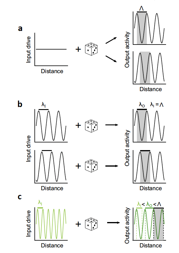
Figure 1. Four predicted properties of a network with local excitation and lateral inhibition: spontaneous periodic patterns, a stable characteristic frequency, phase matching at that frequency, and intermediate frequencies at other stimulation frequencies.
The authors first derived four properties that any network built on local excitation and lateral inhibition should show (Figure 1): (1) it produces periodic patterns of activity on its own, (2) these patterns have a stable characteristic spatial frequency, (3) when stimulated at that frequency, activity locks in phase with the stimulus, and (4) when stimulated at other frequencies, the output settles at an intermediate frequency between the stimulus and the characteristic one.
To test these predictions, ferrets were injected early in postnatal life with the calcium indicator GCaMP6s and the red-shifted opsin ChrimsonR (Figure 2). In immature animals (postnatal days 23–29), the team combined wide-field calcium imaging of the visual cortex with optogenetic stimulation from a 590 nm laser patterned by the Mightex Polygon1000.
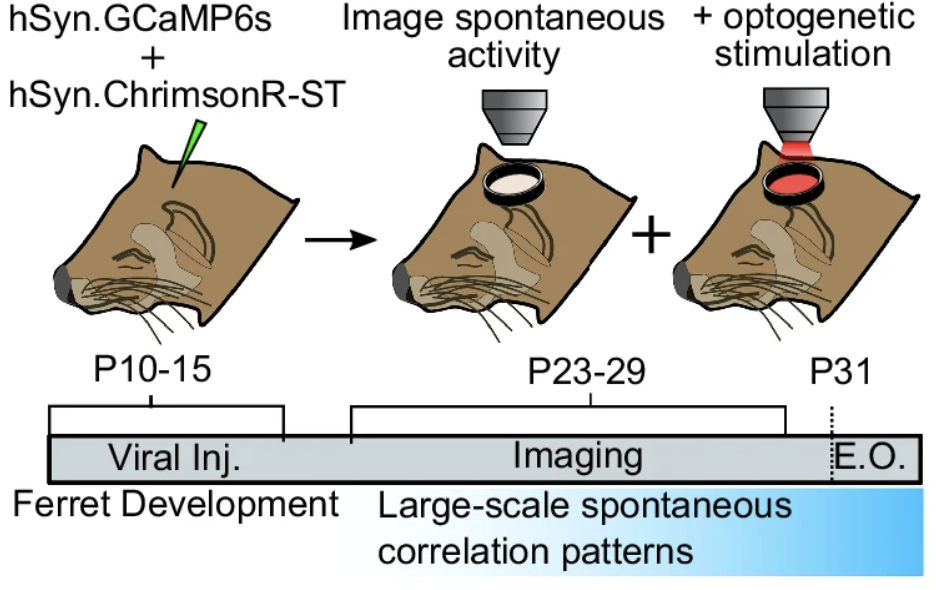
Figure 2. Experimental timeline: GCaMP6s and ChrimsonR were injected in early postnatal life, and combined imaging and stimulation experiments were performed later in postnatal life.
Broad, unstructured stimulation produced clearly modular, periodic activity with a stable characteristic frequency, both within and across animals (Figure 3). When the Polygon projected patterns at each animal’s characteristic frequency, the evoked activity closely matched the phase of the stimulus pattern (Figure 4). Stimulating at other frequencies produced an output frequency that fell between the stimulus and the characteristic frequency (Figure 5).
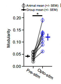
Figure 3. Broad optogenetic stimulation significantly increased the modular organization of visual cortex activity in every animal compared with baseline.
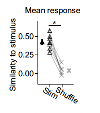
Figure 4. Comparison of stimulation patterns and evoked cortical activity: similarity was high when stimulation matched the tissue’s characteristic frequency, compared with a shuffled control.
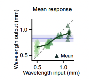
Figure 5. As the stimulation frequency moved away from the characteristic frequency, the output frequency shifted to an intermediate value.
Together, these results show that the immature cortex satisfies all four predictions, supporting the view that local excitation and lateral inhibition are built into cortical circuits early in development and drive the self-organization of modular activity.
All-Optical Interrogation of Millimeter-Scale Networks and Application to Developing Ferret Cortex(Journal of Neuroscience Methods, 403, 110051 (2024))
The neural networks behind sensory perception and behavior often span millimeters of cortex and multiple brain regions. Optical imaging can record activity across these large areas at high spatial resolution, and optogenetics can manipulate specific parts of a network — but combining both at the millimeter scale has been challenging.
To close this gap, the team built an “opto-macroscope” that delivers targeted, patterned optogenetic stimulation across a wide field of view while simultaneously recording calcium activity. A 590 nm laser passes through an acousto-optic modulator for power control and into a Mightex Polygon1000-DL digital micromirror device, which shapes the light into custom stimulation patterns. GCaMP excitation and emission light are routed with dichroic mirrors, and filters keep the stimulation light off the camera (Figure 1).
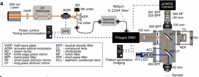
Figure 1. Schematic of the opto-macroscope light path. A 590 nm laser passes through an AOM and into the Polygon DMD for patterned stimulation; GCaMP excitation and emission are routed with dichroic mirrors, and filters block stimulation light from the camera.
Imaging is performed with a back-illuminated sCMOS camera and a tandem-lens macroscope with a 4 mm field of view, and the stimulation patterns are controlled with custom Python software.
Using this system in the developing ferret visual cortex, the authors were able to reactivate endogenous cortical networks, showing that functional modules identified by optical imaging can be directly probed with patterned stimulation. The design’s narrow spectral bandwidth helps reduce photodamage, and it is compatible with a range of red-shifted optogenetic probes, making it a flexible tool for studying neural networks across millimeters of cortex.