



application-banner-mobile-version-flip
Overview

Fluorescence Lifetime Photometry (FLiP) measures the average time a fluorophore remains in an excited state before emitting a photon, typically on a picosecond to nanosecond timescale. Unlike conventional fiber photometry, which measures intensity, FLiP analyzes fluorescence lifetime to deliver highly sensitive quantitative data. This provides deeper insights into biological activity and molecular processes, making it a valuable tool for a wide range of research applications.

What is Fluorescence Lifetime?

Fluorescence lifetime (τ) is the average time a fluorophore remains in its excited state before emitting a photon. Measurements are independent of fluorophore concentration and excitation power, directly reflecting conformational changes and the local microenvironment. Most importantly, it allows for sensitive detection of subtle lifetime changes, providing a stable and specific quantitative readout for in vivo studies.

application-page-schematic-flip-what-is-flourescence-lifetime
Key Benefits of Fluorescence Photometry

Fluorescence lifetime provides a quantitative and reliable approach to monitoring fluctuations in biosensor activity. Lifetime measurements provide significant experimental advantages. It allows for long-term, stable and sensitive recordings. By recording absolute measurements, researchers can mitigate unintended inter-subject variability, establish event-independent baselines, and maximize signal-to-noise ratio.

ENABLES

Quantitative & Absolute Measurements
Obtain consistent measurements across different samples and conditions, eradicating baseline normalizations and intensity-related variability.

Reliable Long-Term Monitoring
Stable fluorescence measurements over extended recording periods, making FLiP particularly useful for longitudinal and continuous studies.

Sensitive Detection of Biological Changes
Detection of subtle changes in molecular interactions and analyte levels using lifetime-sensitive probes.

MINIMIZES

Variability Related to Probe Expression
Differences in probe expression leading to variations in fluorescence intensity.

Photobleaching-Related Artifacts
Reductions in the magnitude of event-related signals over the course of prolonged illumination.

Motion-Related Artifacts
Intensity artifacts caused by movement of the sample or animal altering collection of fluorescence signal.

Diverse Research Applications

Quantitative Monitoring of Brain Signaling

fluorescence lifetime photometry allows for Quantifying tonic and phasic changes in brain signaling using calibrated fluorescence lifetime measurements and compatible biosensors.

Quantify tonic and phasic changes in brain signaling using calibrated fluorescence lifetime measurements and compatible biosensors.

Robust, Long Term Experiments
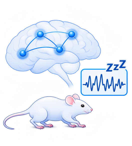
fluorescence lifetime photometry allows for High confidence, multi-time point, and long duration experiments. Relevant for research areas including development and sleep.

High confidence, multi-time point, and long duration experiments. Relevant for research areas including development and sleep.

Study Neurological Diseases & Disorders

fluorescence lifetime photometry allows Investigation of changes in neural signaling between healthy and diseased models through stable, long-term fluorescence recordings.

Investigate changes in neural signaling between healthy and diseased models through stable, long-term fluorescence recordings.

Diverse Biosensors
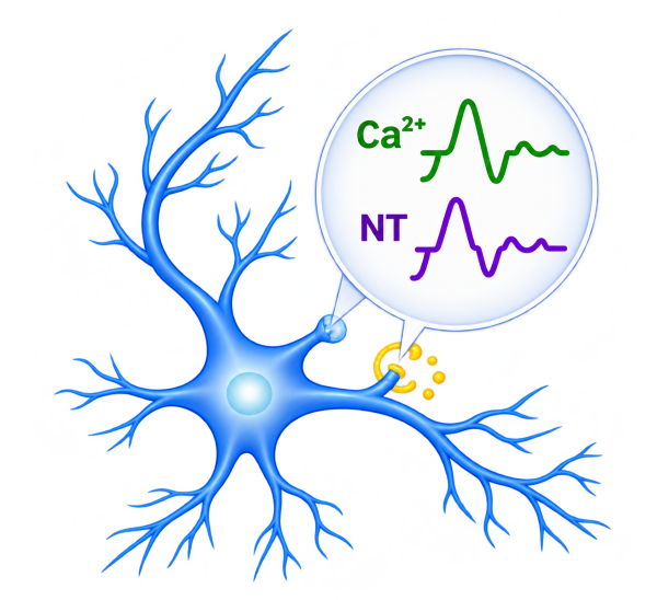
fluorescence lifetime photometry allows for Measuring changes in calcium and neurotransmitter signaling using compatible fluorescent indicators and emerging lifetime-sensitive biosensors.

Measure changes in calcium and neurotransmitter signaling using compatible fluorescent indicators and emerging lifetime-sensitive biosensors.

OASIS FLiP at a Glance

Fluorescence lifetime provides a quantitative and reliable approach to monitoring fluctuations in biosensor activity. Lifetime measurements provide significant experimental advantages. It allows for long-term, stable and sensitive recordings. By recording absolute measurements, researchers can mitigate unintended inter-subject variability, establish event-independent baselines, and maximize signal-to-noise ratio.
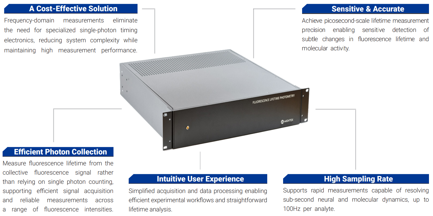
OASIS FLiP labelled system at a glance

Tap image to enlarge

OASIS FLiP enables robust measurements of calcium dynamics, neurotransmitter signaling, and molecular activity in vivo. It supports reliable, long-term monitoring of neural activity, providing quantitative measurements and a stable baseline while mitigating photobleaching- and motion-related artifacts. This benefits a wide range of basic and translational behavioural neuroscience research areas.

System Specifications
Excitation Wavelength OptionsCustomizable
Measurement Precision50 ps (ms)
Lifetime Range1-10 ns
Sampling Rateup to 100 Hz
DIO Countup to 12
Voltage Level3.3-5 V
Optical Fiber Core Diameter200 µm or 400 µm
NA0.39
SubstrateLifetime BiosensorExcitation Wavelength
Ca2+Tq–Ca–FLITS 1439 nm
tr-CCaMP 2437 nm
tr-GCaMP 2493 nm
tr-RCaMP 2563 nm
RCaMP1h 3560 nm
LifeCaMP 4488 nm
AChGRAB ACh3.0 5488 nm
DAdLight3.8 6488 nm
PKAtAKARα 7488 nm
tAKARβ 7488 nm
tAKARγ 7488 nm
FLIM-AKAR 8488 nm
PKCCKAR3 9488 nm
Extracellular GlutamateRncp-iGluSnFR1 10560 nm
SerotoninsDarken 11488 nm
GlucoseiGlucoSnFR-TS 12400 nm
LactateLiLac 13425 nm
ATPqMaLioffG 14488 nm
  1. van der Linden, F. H. et al. A turquoise fluorescence lifetime-based biosensor for quantitative imaging of intracellular calcium. Nat. Commun. 12, 7159 (2021).
  2. Tan, Z. et al. Time-resolved genetically encoded indicators toward quantitative imaging of calcium dynamics in living cells. ACS Sens. 11, 2142–2152 (2026).
  3. Díaz-García, C. M. et al. Neuronal stimulation triggers neuronal glycolysis and not lactate uptake. Cell Metab. 26, 361–374.e4 (2017).
  4. Lodder, B. et al. Rapid fluorescence lifetime sensor development of LifeCamp enables transient and baseline absolute calcium measurements. bioRxiv (2025) doi:10.64898/2025.12.23.696288.
  5. Ma, P. et al. Fast and slow: Recording neuromodulator dynamics across both transient and chronic time scales. Sci. Adv. 10, eadi0643 (2024).
  6. Roshgadol, J. I. et al. Sensitive dLight3 for imaging broad-spectrum dopamine events across brain regions. Research Square (2025).
  7. Ma, L. et al. A highly sensitive A-kinase activity reporter for imaging neuromodulatory events in awake mice. Neuron 99, 665–679.e5 (2018).
  8. Tilden, E. I. et al. A Cre-dependent reporter mouse for quantitative real-time imaging of protein kinase A activity dynamics. Sci. Rep. 14, 3054 (2024).
  9. Yahiro, T. et al. A high-performance genetically encoded sensor for cellular imaging of PKC activity in vivo. Nat. Commun. 16, 6378 (2025).
  10. Zhou, H.-M. et al. Quantification of extracellular glutamate levels using fluorescence lifetime imaging of the red genetically encoded biosensor Rncp-iGluSnFR1. Microchem. J. 212, 113571 (2025).
  11. Kubitschke, M., Beck, V. & Masseck, O. A. Fluorescence lifetime imaging of sDarken as a tool for the evaluation of serotonin levels. bioRxiv 2024.01.04.574197 (2024).
  12. Díaz-García, C. M. et al. Quantitative in vivo imaging of neuronal glucose concentrations with a genetically encoded fluorescence lifetime sensor. J. Neurosci. Res. 97, 946–960 (2019).
  13. Koveal, D. et al. A high-throughput multiparameter screen for accelerated development and optimization of soluble genetically encoded fluorescent biosensors. Nat. Commun. 13, 2919 (2022).
  14. Arai, S. et al. qMaLioffG: A single green fluorescent protein FLIM indicator enabling quantitative imaging of endogenous ATP. Research Square (2023) preprint, doi:10.21203/rs.3.rs-3343438/v1.

Contact Us

This field is for validation purposes and should be left unchanged.
Mightex will only use your personal information to provide the products and services you have requested. You may unsubscribe at any time. We value your privacy.
To learn more about unsubscribing, our privacy protocols, and the measures we take to ensure your privacy, we invite you to explore our comprehensive Privacy Policy.