



Investigating Somatostatin Neuron Activity in Freely Moving Mice with the OASIS Fiberscope and Photometry

Huanhuan Li is a researcher in Prof. Geoffrey Lau’s lab at City University of Hong Kong. His research combined in vivo calcium imaging and optogenetics to investigate how somatostatin-expressing neurons influence social behavior, stress, and depression-related behaviors in freely moving mice. His work used Mightex’s OASIS Fiberscope system to visualize neural activity with cellular and spatio-temporal resolution.
Research Highlights
In Vivo Calcium Imaging of Somatostatin-Expressing Cell Activity in the Cingulate Cortex During Social Interaction in Freely Moving Mice Using Mightex’s OASIS Fiberscope(Mightex Research Excellence Award, 2021)
Somatostatin (SST)-expressing neurons are a powerful inhibitory cell type distributed throughout the limbic system, where they help shape circuits underlying motivation, mood, and social behavior. Huanhuan Li and colleagues in Prof. Geoffrey Lau’s lab at the City University of Hong Kong set out to understand how this single class of inhibitory neuron behaves across two different limbic regions and two very different behavioral contexts — using the Mightex OASIS system to record SST activity in freely moving mice with cellular and spatio-temporal resolution.
The cingulate cortex (CC) is a key brain region in the limbic system that coordinates actions and motivational behaviors. Somatostatin-expressing GABAergic neurons in the cingulate cortex (CCSST) can provide powerful inhibition to the CC circuitry through high basal firing activity and synchronized firing [1]. An open question is how CCSST cells contribute to motivational behaviors such as sociality.
Our lab is particularly interested in understanding the dynamic changes of inhibitory neurons in response to reward as well as stress. We found that a subset of CCSST cells was recruited upon social stimuli in the three-chambered social interaction test (Figure 1A, B).

Figure 1. Experimental Overview.
Their first target was the cingulate cortex (CC), a region that helps coordinate motivated behavior. Somatostatin-expressing GABAergic neurons there (CCSST) provide strong inhibitory tone through high basal firing and synchronized activity, but whether CCSST cells played a direct role in social motivation was still an open question. Using the Mightex OASIS Fiberscope system in a three-chambered social interaction test, the team recorded Ca2+ activity from CCSST cells while two male mice interacted freely in an open field, tracking interaction time and inter-mouse distance alongside neural activity in real time.
A subset of CCSST cells responded specifically to social stimuli: activity rose whenever the resident and intruder mice came into close proximity, staying elevated through both passive and active bouts of interaction.
To visualize CCSST neuronal activation in the spatio-temporal and cellular resolutions, we measured the Ca2+ activity of CCSST cells by incorporating the Mightex OASIS Fiberscope system into our behavioral setup (Figure 1C). To examine the relationship between social interaction and CC neural activity, we placed two male mice in an open field. We measured the time spent by the mice interacting with each other as well as the distance between them, while recording the response simultaneously (Video 1). In the reciprocal social interaction test, CCSST neuronal activity was persistently elevated during both passive and active social interactions — CCSST neurons were activated when the intruder and resident mice were in close distance (Figure 1D).
Video 1. Calcium imaging data of CCSST neurons and corresponding social interaction behaviour.
We will further characterize the role of CCSST cells in these behaviors by combining the Mightex OASIS Fiberscope system and cell-type-specific manipulation tools. In sum, the Mightex OASIS system provides a powerful tool for investigating the relationship between neural population activity and social behavior in real-time.
[1] Urban-Ciecko J, Barth AL. Somatostatin-expressing neurons in cortical networks. Nat Rev Neurosci. 2016 Jul;17(7):401-9.
This pilot dataset, which earned 2nd place in the Mightex Research Excellence Award, showed that CCSST neurons track social proximity in real time. It also raised a natural next question: does SST-expressing activity play an equally active role in other limbic regions and other behavioral states — like stress and mood?
OASIS Photometry Publication Feature: Activation of Somatostatin-Expressing Neurons in the Lateral Septum Improves Stress-Induced Depressive-like Behaviors in Mice(Pharmaceutics, 14(10), 2253 (2022))
Check out this recent publication using the OASIS Photometry! The paper outlines research by Huanhuan Li and colleagues from Geoff Lau’s lab at the City University of Hong Kong.
Introduction
The limbic system, including the lateral septum (LS), plays a crucial role in mood disorders, such as depression. However, the role of the LS in depression remains unclear. A potential important role is ascribed to neurons expressing somatostatin (SST) in particular. Li and colleagues hypothesized that LS SST neuronal activity is linked to stress and depression.
Methods & Findings
In order to examine the contributions of SST-expressing neurons of the LS in depression-like behaviors in mice, Li and colleagues leveraged calcium imaging using the Mightex OASIS Photometry and GCaMP6s sensor in SST-cre mice. Their findings show that LS SST neuronal activity was significantly reduced in two mouse models of depression (Chronic Restraint Stress [CRS] and repeated lipopolysaccharide [LPS] injections).
In order to determine whether artificial manipulation of LS SST neuronal activity could attenuate the depression-like behavioural observations, Li and colleagues combined their calcium imaging with optogenetics to excite LS SST neurons during depression model experiments. The authors found that optogenetic excitation of LS SST neurons produced antidepressant-like effects in the LPS depression model. Taken together these findings elucidate the LS SST neurons as a potential therapeutic target for the treatment of depression.
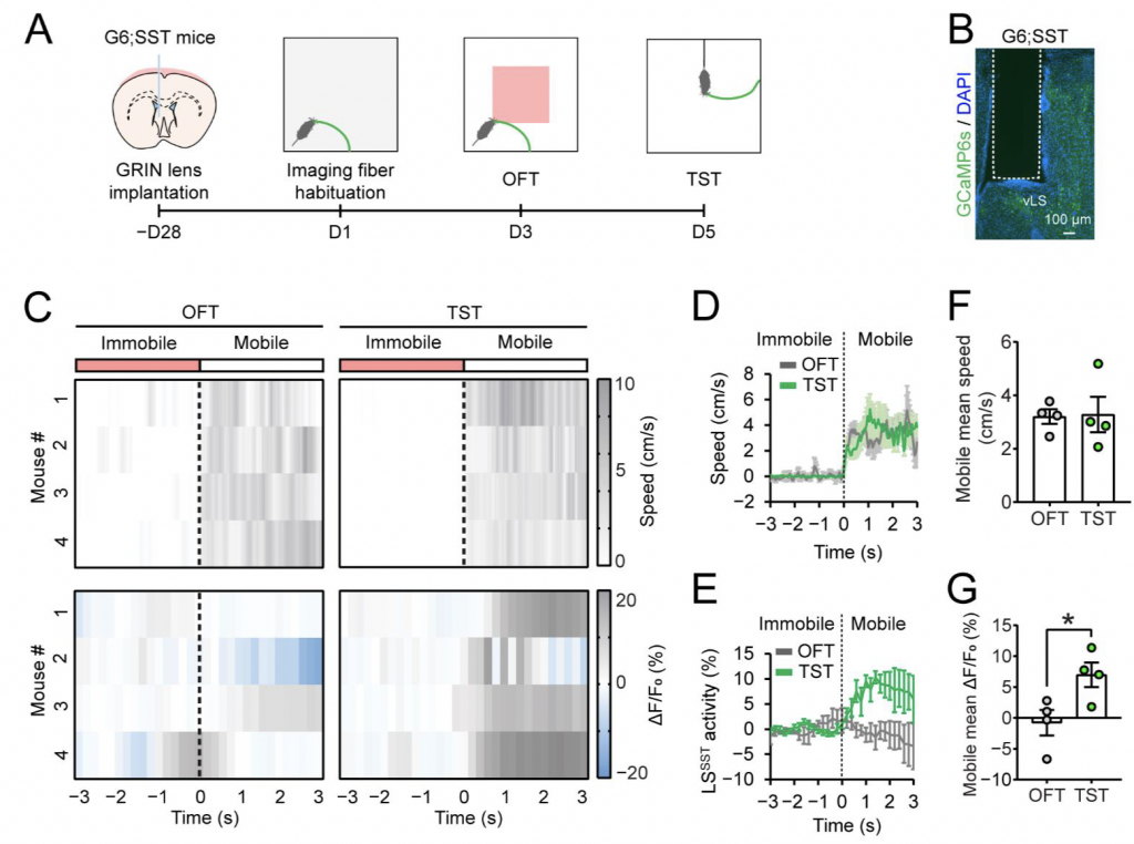
Figure 1: Adapted from Source
Panel A. The surgical and experimental preparation stages.
Panel B. GRIN lens location within LS.
Panel C. Calcium imaging results. Change in fluorescence depicted for 4 mice; changing in fluorescence (bottom) and corresponding behavioural changes (top) seen as gradient overtime for Open Field Test (OFT; left) and Tail Suspension Test (TST; Right)
Panels D & F. Behavioural quantification for OFT and TST; the dotted line represents the initiation of the mobile phase.
Panels E & G. Calcium imaging quantification for LS SST neurons during OFT and TST; the dotted line represents the initiation of the mobile phase.
Findings and Conclusion
The above described experiments demonstrate the capabilities of the Mightex OASIS Photometry for calcium imaging in freely moving mice. To learn more about using the system for your research, get in touch today! To read the full publication, please click here.