



Exploring Cellular Signaling in Learning and Memory with the OASIS Fiberscope

Jayant Rai is a PhD researcher in the Department of Molecular Genetics at the University of Toronto, working with Prof. Kenichi Okamoto at the Lunenfeld-Tanenbaum Research Institute and Dr. Mei Zhen. His research combines genetic, pharmacological and optogenetic manipulation of intracellular signaling, in particular cAMP, with in vivo calcium imaging of hippocampal neurons to understand how signaling shapes circuit activity during learning and memory in freely moving mice. His work uses Mightex’s OASIS Fiberscope system.
Research Highlights
Elucidating Dynamic Intracellular Signalling Pathways of Hippocampus-Dependent Memory with the OASIS Fiberscope(Mightex Research Excellence Award, 2021)
Understanding how neural circuits process memory information is essential for elucidating the mechanisms underlying learning and memory. Previous studies have demonstrated that hippocampal neurons display highly repetitive patterns of synchronous activity that are involved in learning and memory. However, the molecular mechanisms underlying the coordination of circuit activity that serve for learning and memory remain unclear.
By combination of genetic, pharmacological and optogenetic manipulation of intracellular signalling with in vivo live calcium imaging of hippocampal neurons in freely behaving mice using Mightex OASIS Fiberscope micro-endoscopy system, we aim to address dynamic intracellular signaling mechanisms that regulate neuronal circuit activity during hippocampus-dependent memory. Attached is a video demonstrating in vivo GCaMP6 fluorescence imaging in hippocampal CA1 pyramidal neurons with the Mightex OASIS Fiberscope micro-endoscopy system (Video 1).
Video 1. Calcium imaging data from the hippocampal CA1 neurons.
Video 2. Corresponding freely-behaving mouse in an Open Field Test.
Using this system, we can characterize the activity pattern of CA1 neurons across different stages of hippocampus-dependent memory (formation, consolidation, recall) in freely-behaving mice during behaviour assays (as shown by the Open Field Test in Video 2). Future planned experiments in our lab will utilize the Mightex OASIS Fiberscope system to conduct simultaneous optogenetic stimulation of neuronal activity while observing calcium dynamics and mouse behaviour in the same animal during memory tests.
We are able to record calcium imaging and mouse behaviour data in the same animal and currently I am working to connect our calcium imaging data to the mouse behaviour (i.e., to identify patterns of neuronal activity during specific behaviours such as interacting with a novel object).
Optical Interrogation of cAMP Signaling in Behaving Animals Reveals cAMP-Dependent Neural Circuit Activity Underlying Hippocampus-Dependent Short-Term Learning and Memory(Mightex Travel Award, SfN 2025)
The spatiotemporal organization of hippocampal circuit activity is critical for hippocampus-dependent learning and memory. However, the molecular mechanisms underlying the coordination of hippocampal neural activity and their task-related dynamics remain unclear. Here, we investigate the role of cAMP (adenosine 3′,5′-cyclic monophosphate) signaling to regulate CA1 neural activity during hippocampus-dependent short-term (STM) by optogenetic cAMP manipulation and fiber-based calcium imaging in freely-behaving animals.
cAMP is a ubiquitous second messenger in various intracellular signaling pathways that mediate sensory transduction and neuromodulation and serves for certain types of synaptic plasticity. We previously reported the spatiotemporal function of cAMP to rapidly enhance synaptic strength and neuronal depolarization in murine hippocampal slices, suggesting that cAMP may not only serve synaptic potentiation but may also regulate neural circuit activity during memory.
To test this function, we virally expressed light-sensitive enzymes PAC (Photoactivatable Adenylyl Cyclase) or light activatable Dr-HsPDE4 (Phosphodiesterase 4) in CA1 neurons for light dependent cAMP enhancement or suppression, respectively, by activation of a chronically implanted wireless fiber- optic LED in freely-behaving mice. During object recognition memory tests, we observe a deficit in object memory following cAMP suppression specifically during training, while cAMP enhancement during training promotes STM, indicating a cAMP function that is critical for STM formation.
To directly observe the role of cAMP in hippocampal CA1 neural circuit activity during STM formation, we virally coexpress a calcium indicator (GCaMP8m) with cAMP metabolic enzymes, then implanted a GRIN lens coupled with the wireless fiber optic LED by our custom headmount system. We optically recorded activity of CA1 neurons with or without cAMP manipulation by micro-endoscopic calcium imaging in freely-behaving mice during STM tests, then fit a latent space dynamical system model to estimate population dynamics underlying memory task-specific behaviors. We observed that cAMP perturbation disrupts the task-related structure and dynamics in the latent embedding of population activity during STM, which correlates with observed deficit in memory behaviour by cAMP manipulation.
Thus, our results reveal that the spatiotemporal cAMP signal is critical for CA1 activity that facilitates hippocampus-dependent memory. We will further discuss our approach to characterize the cAMP mechanism of action to regulate population dynamics during hippocampus-dependent STM and LTM.

Investigating the Role of cAMP as a Critical Regulator of CA1 Neuronal Population Activity in Mice During Hippocampus-Dependent Spatial Learning & Memory(Mightex Travel Award, SfN 2025)
The spatiotemporal organization of neuronal population activity is critical for hippocampus-dependent learning & memory. However, the molecular mechanisms underlying the coordination of CA1 neuronal activity and their task-related dynamics remain unclear. Here, we investigate the role of cAMP (adenosine 3′,5′-cyclic monophosphate) signaling to regulate CA1 neuronal activity during hippocampus-dependent spatial learning & memory by cAMP manipulation and fiber-based calcium imaging in freely-behaving mice. cAMP is a ubiquitous secondary messenger that mediates sensory transduction and neuromodulation.
We previously reported a rapid cAMP function to enhance the spatial propagation of depolarizing field potentials across murine hippocampal slices, suggesting cAMP may serve to enhance neuronal population activity for memory. To characterize the role of cAMP for hippocampus-dependent spatial learning & memory, we virally expressed the light-sensitive enzyme PAC (Photoactivatable Adenylyl Cyclase) or constitutively active enzyme PDE4cat (Phosphodiesterase 4) in CA1 neurons in freely-behaving mice for cAMP enhancement or suppression, respectively, during the object location test (OLT) and contextual fear conditioning (CFC) test. In the OLT, we observe that cAMP suppression results in a significant memory deficit while cAMP enhancement during training (but not testing) promotes the displaced object preference, indicating a cAMP function critical for encoding of short-term spatial recognition memory.
In the CFC test, we utilize a Dox inducible activity-dependent expression system to specifically express PAC or PDE4cat in CA1 neurons activated during CFC. After 24h, we assayed the effect of cAMP manipulation during exposure to the spatial environment (context). We found a significant reduction in freezing behaviour with cAMP suppression compared to control, suggesting that cAMP is critical for context-dependent fear memory retrieval. To directly examine how cAMP may affect CA1 population activity during OLT and CFC test, we recorded the CA1 neuronal response profiles by in vivo calcium imaging in freely-behaving mice. We found similar frequency and duration of neuronal calcium activities between the control and cAMP suppression groups, however, the organization of latent neuronal population states was perturbed by cAMP suppression, suggesting that cAMP may regulate CA1 population activity underlying spatial memory. Further, we will discuss our approach for combined calcium imaging and cAMP optogenetics in the same animal to directly interrogate the effect of cAMP perturbation on CA1 population dynamics during learning & memory.
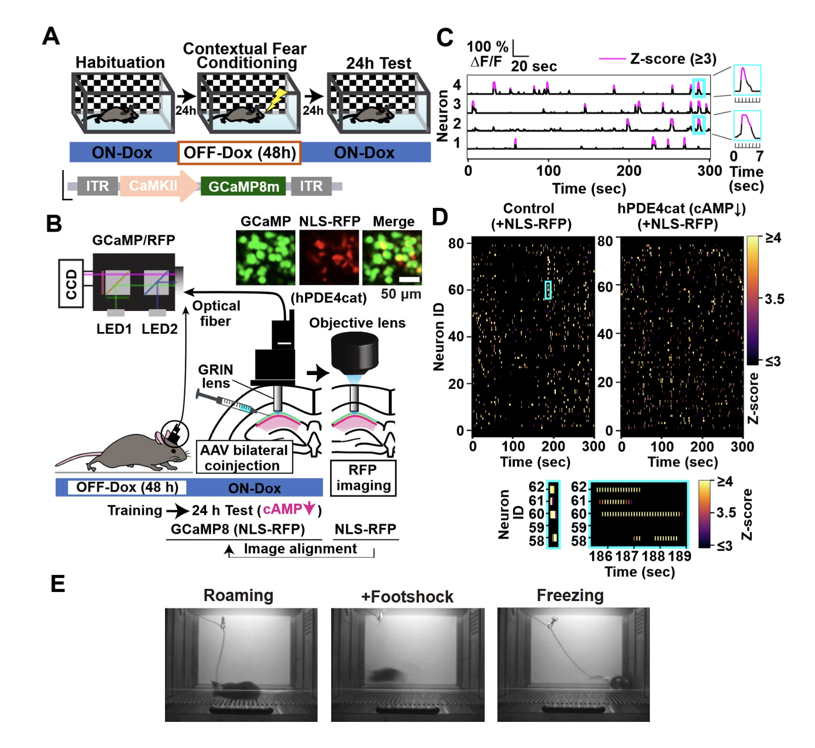
Figure 1. In vivo fiber-based calcium imaging during contextual fear conditioning using the Mightex OASIS Fiberscope system.
(A) Schematic for the contextual fear conditioning (CFC) assay. GCaMP8m was virally expressed in CA1 neurons under a CaMKIIα promoter. Dox-inducible activity-dependent expression of a mutant phosphodiesterase was used for cAMP suppression in CA1 neurons activated during CFC.
(B) A schematic of in vivo Ca2+ imaging during contextual fear memory retrieval test.
(C) Sample traces of in vivo Ca2+ activity during contextual fear memory retrieval test. The red line indicates the threshold for calcium activity (≥ 3 z-score).
(D) Top: Heatmap for sample activities of the NLS-RFP positive neurons (≥ 3 z-score) across time during contextual fear memory retrieval test in a control and cAMP suppression mice. Bottom: Expanded plot from the blue square showing z-score traces over a 3-second window obtained by 10 Hz imaging.
(E) Representative images of mouse behaviors during the CFC assay.